
Figures & data
Figure 1 A case with a small choroidal nevus in the posterior pole of the left eye with some small drusen over the lesion.
Notes: (A) The fundus photograph of a pigmented lesion on the macular area without exudative changes. (B) B-scan showing a nearly flat, small choroidal lesion with medium-to-high internal reflectivity. (C) OCTA with slabs crossing the surface of the choroidal nevus at the choriocapillaris level by en face imaging with intact outer retina (arrow). (D) The flow rate over the lesion at the level of choriocapillaris level was evaluated (arrow). It elucidates more concentrated vasculature but is still not much different than nearby retina.
Abbreviation: OCTA, optical coherence tomography angiography.
Abbreviation: OCTA, optical coherence tomography angiography.
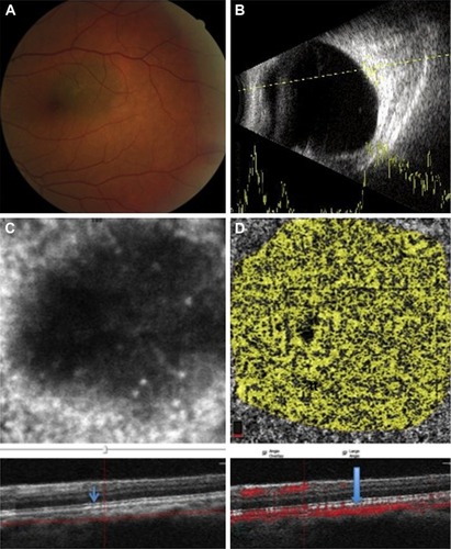
Figure 2 A case with a small and growing peripapillary choroidal melanoma.
Notes: (A) The fundus photograph demonstrates a pigmented superior peripapillary lesion and pigmentary changes in the macular area due to multiple episodes of increasing subretinal fluid. (B) B-scan shows a small choroidal lesion with medium-to-high internal reflectivity. (C) Cross-sectional B-scan OCTA with slabs crossing the surface of the choroidal melanoma through the choriocapillaris. The RPE–Bruch’s membrane complex shows some derangement with subretinal fluid (arrow) on en face imaging. (D) The flow rate over the lesion at the level of choriocapillaris level was measured, showing thinner vascular area than the nearby choriocapillaris (arrow).
Abbreviations: OCTA, optical coherence tomography angiography; RPE, retinal pigment epithelium.
Abbreviations: OCTA, optical coherence tomography angiography; RPE, retinal pigment epithelium.
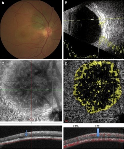
Table 1 Demographic findings of the patients with small melanocytic nevi and treatment-naive small choroidal melanomas
Table 2 OCTA features of the patients with small melanocytic nevi and treatment-naive small choroidal melanomas
Table 3 OCTA FAZ and flow rate in the affected eye with melanoma or nevus versus the fellow eye in 11 consecutive cases