Figures & data
Figure 1 Coronal A) and sagittal B) computed tomography images showing a large fracture of the left orbital floor with herniation of a large volume of orbital fat and a vertically elongated inferior rectus muscle extending to the fracture site.
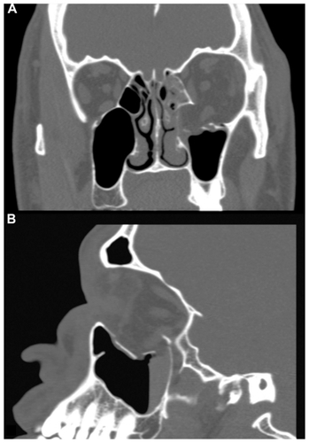
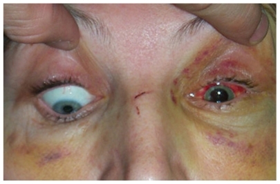
Figure 2 A patient with a right medial orbital wall fracture and medial rectus entrapment demonstrates restriction of abduction of the right eye.
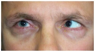
Figure 3 A patient with a left orbital floor fracture and inferior rectus entrapment demonstrates restriction of downgaze of the left eye.