
Figures & data
Figure 1 Color fundus photo and optical coherence tomography, showing optic disk pit (white arrows) and macular elevation.
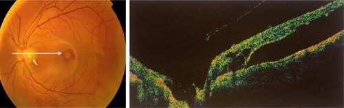
Figure 2 Optic disc pit in a male patient.
Notes: Fluorescein angiography and optical coherence tomography, showing optic disk pit at the temporal side of the disk with macular elevation (upper panel). Fundus photo and optical coherence tomography of the same patient 12 years after macular buckling procedure, where there is absorption of the fluid (lower panel). A triangular-shaped space at the right side of the optic nerve is evident, and the lack of communication between this triangular space and the intraretinal layers is probably due to the sponge fixation at the nasal side of the optic nerve. White arrows represent the macular elevation.
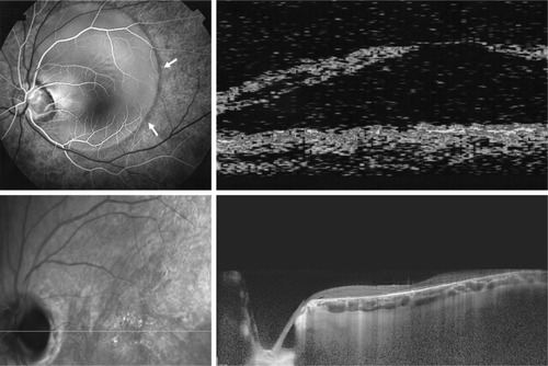
Table 1 Main treatment modalities for optic disk pit maculopathy and their anatomical and functional results