Figures & data
Figure 1 Two patterns of RRD based on the presence of PVD.
Abbreviations: PVD, posterior vitreous detachment; RRD, rhegmatogenous retinal detachment; SRF, subretinal fluid.
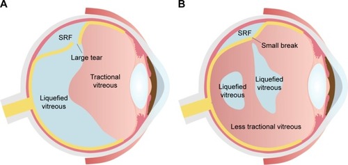
Figure 2 Different patterns of a retinal break in rhegmatogenous retinal detachment associated with PVD.
Abbreviation: PVD, posterior vitreous detachment.
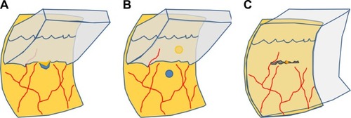
Figure 3 Chronic rhegmatogenous retinal detachment with subretinal strand.
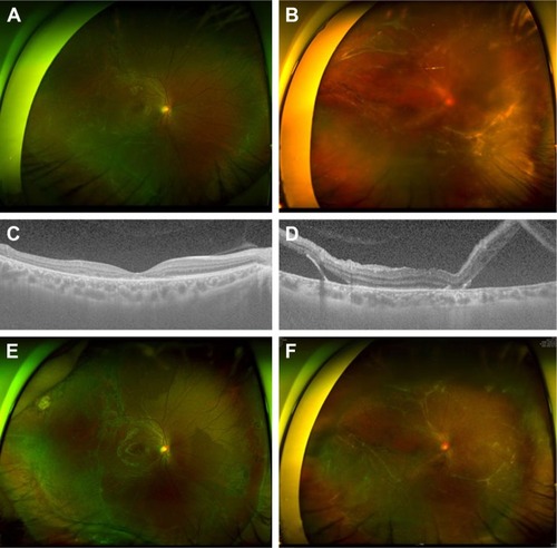
Figure 4 Different effects of SB according to the vitreous status.
Abbreviations: PVD, posterior vitreous detachment; RRD, rhegmatogenous retinal detachment; SB, scleral buckling.
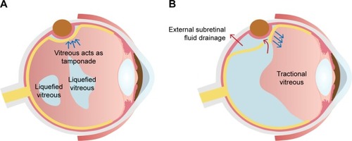
Figure 5 Intraocular gas tamponade can be an effective adjuvant for scleral buckling when external drainage of SRF fails to close the break because of the extensively liquefied vitreous.
Abbreviation: SRF, subretinal fluid.
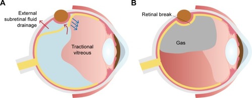
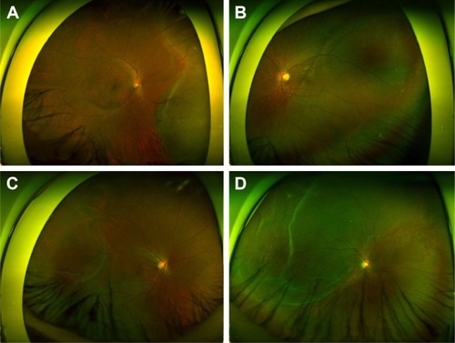
Figure 6 A case of RRD with fluidic vitreous humor without PVD. A 12-year-old girl presented with visual loss for 3 days.
Abbreviations: PVD, posterior vitreous detachment; RRD, rhegmatogenous retinal detachment.
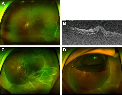
Figure 7 Cases of rhegmatogenous retinal detachment less suitable for SB alone. PVD, bullous retinal detachment, and a relatively large retinal tear are noted.
Abbreviations: PVD, posterior vitreous detachment; SB, scleral buckling.
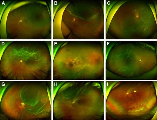
Figure 8 Cases that are good candidates for scleral buckling.