
Figures & data
Table 1 Demography and clinical characteristics of eyes undergoing pars plana vitrectomy
Table 2 Comparison of the mean retinal thickness of postoperative eyes after macular hole surgery at 6 months and corresponding fellow eyes (µm, mean ± SD)
Figure 1 SD-OCT images of a 71-year-old man with a stage 2 MH who underwent PPV with peeling of the ILM.
Abbreviations: BCVA, best-corrected visual acuity; GCC, ganglion cell complex; ILM, internal limiting membrane; MH, macular hole; PPV, pars plana vitrectomy; RPE, retinal pigment epithelium; SD-OCT, spectral-domain optical coherence tomography.
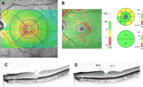
Figure 2 SD-OCT images of a 62-year-old woman with stage 3 MH after PPV without peeling of the ILM.
Abbreviations: BCVA, best-corrected visual acuity; GCC, ganglion cell complex; ILM, internal limiting membrane; MH, macular hole; PPV, pars plana vitrectomy; RPE, retinal pigment epithelium; SD-OCT, spectral-domain optical coherence tomography.
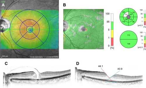
Table 3 Comparison of the mean retinal thickness of postoperative eyes after macular hole surgery at 6 months with and without ILM peeling (µm, mean ± SD)
Table 4 Comparison of the mean parafoveal GCC of postoperative eyes after macular hole surgery with ILM peeling and without ILM peeling (µm, mean ± SD)
Table 5 Comparison of the mean retinal slope of postoperative eyes after macular hole surgery with ILM peeling and without ILM peeling (°, mean ± SD)