Figures & data
Figure 1 An OWC measuring 3 cm in diameter has been developed for ocular use with a flexible base that attaches to the perimeter of the eye (A). The device has a transparent cover and a self-sealing port that allows for the delivery of therapeutics (A). The OWC has been specifically designed for use in our guinea pig model (B) and provides a watertight seal that allows for the creation of a controlled microenvironment over the eye. The base, cover, and self-sealing port are labeled accordingly as indicated by arrows (B).
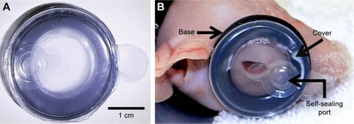
Table 1 Pathology scoring system
Figure 2 Re-epithelialization of corneal wounds. Representative images of white light (A) and fluorescein stained (B) injured eyes at 0, 24, 48, and 72 hours. Comparison of percentage wound closure between groups over time (C). No statistically significant differences across treatment groups. N=5 animals per group.
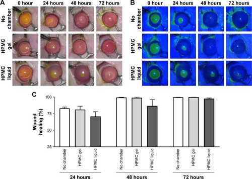
Figure 3 Corneal thickness. OCT of control, no chamber, HPMC gel- and liquid-treated guinea pig corneas at 0, 24, 48, and 72 hours (A). Comparison of central corneal thickness between groups over time (B). N=5 animals per group. *P<0.05; **P<0.005.
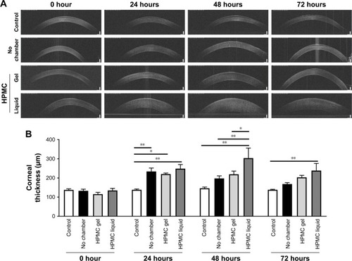
Figure 4 IOP of treated and untreated guinea pig eyes at 0, 24, 48, and 72 hours. No statistically significant differences across treatment groups. N=5 animals per group.
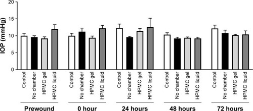
Figure 5 Histological analysis of guinea pig corneas. Representative images of hematoxylin and eosin stained cornea sections (A). Comparison of key pathology observations between treatment groups (B). Comparison of total number of epithelial mitotic figures per group (C). No clinically relevant findings or any significant differences were observed.
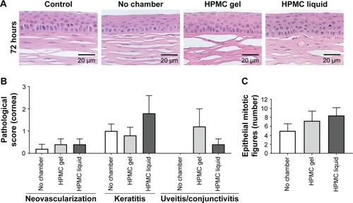
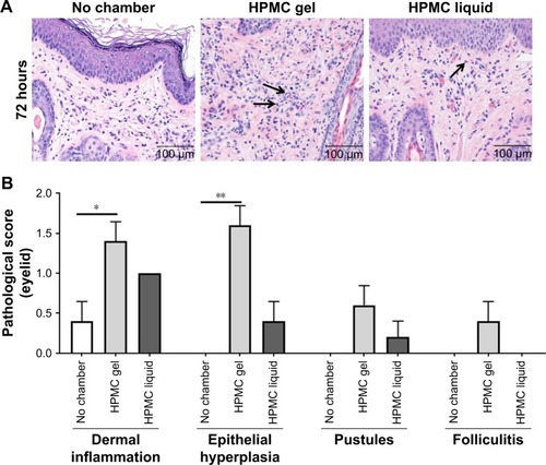
Figure 6 Histological analysis of guinea pig eyelid skin. Representative images of hematoxylin and eosin stained skin sections (A). Comparison of key pathology observations between treatment groups (B). Arrows indicate heterophilic inflammatory cells. Significant differences in dermal inflammation and epithelial hyperplasia were observed in scored eyelid sections between the HPMC gel treated groups and untreated controls. *P<0.05; **P<0.005.