
Figures & data
Figure 1 Both the endoscopic image (left) and the wide-angle viewing image (right) are integrated on the large 3D monitor that is placed at the foot of the bed. The small conventional monitor for the endoscope is placed on the left side of the surgeon.
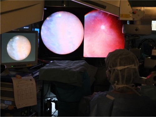
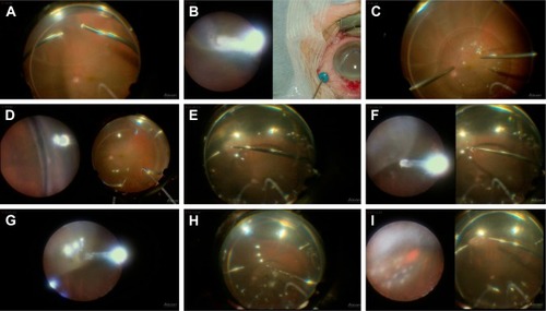
Figure 2 Intraoperative 2D snapshots in a case of hybrid surgery for rhegmatogenous retinal detachment. (A) Core vitrectomy performed using a wide-angle view (WAV). (B) Cryopexy to fix a tiny retinal break identified intraoperatively at the ora serrata using the endoscopic view (left). A microscopic view of the outer segment is simultaneously visualized (right) and helps to prevent freezing of the cornea or the lid. (C) Injection of perfluorocarbon liquid (PFCL) while using WAV. (D) Peripheral vitreous shaving performed while using the endoscopic view without any scleral depression. (E) Fluid/air exchange and removal of PFCL using WAV. (F) Internal drainage through an original retinal break at the periphery. The images from both the endoscopic view (left) and WAV (right) are integrated on the same monitor. (G) Endoscopic view of the internal drainage. (H) PFCL is completely removed while using WAV. (I) Laser photocoagulation of a retinal break while using the magnified endoscopic view (left). Simultaneous visualization by WAV (right) helps with the orientation of the endoscopic manipulation.
Video S1 shows intraoperative side-by-side stereo image in a case of hybrid surgery for rhegmatogenous retinal detachment (same case as shown in ). Core vitrectomy was performed while using a wide-angle view (WAV). Cryopexy was applied to a tiny retinal break identified intraoperatively at the ora serrata when using the endoscopic view (left). Simultaneous visualization of the microscopic view for the outer segment (right) helps to prevent freezing the cornea or the lid. Perfluorocarbon liquid (PFCL) was injected while using WAV. Peripheral vitreous shaving was performed using the endoscopic view without any scleral depression. Fluid/air exchange and removal of PFCL was performed while using WAV, followed by internal drainage through an original retinal break at the periphery. Images from both the endoscopic view (left) and WAV (right) are integrated on the same monitor. Also, the endoscopic view alone can be monitored. PFCL is completely removed while using WAV. Laser photocoagulation of a retinal break while using the magnified endoscopic view (left). Simultaneous visualization of the WAV helps with orientation of the endoscopic manipulation.