
Figures & data
Figure 1 Illustration of the 4 criteria of severity on FA and OCT.
Notes: The 4 criteria are as follows: 1) cumulative areas of >5 DD of diffuse atrophic retinal pigment epithelium alterations as visualized on mid-phase FA (A). 2) Multiple (at least 2) “hot spots” of leakage separated by at least 1 DD of non-hyperfluorescent healthy-appearing retina on mid-phase FA (B). 3) An area of diffuse fluorescein leakage with a surface of >1 DD on mid-phase FA, without an evident leaking focus (C). 4) Presence of posterior cystoid retinal degeneration on OCT (D and E).
Abbreviations: DD, optic disc diameters; FA, fluorescein angiography; OCT, optical coherence tomography.
Abbreviations: DD, optic disc diameters; FA, fluorescein angiography; OCT, optical coherence tomography.
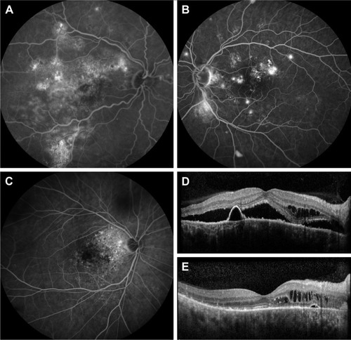
Table 1 Patient demographics
Figure 2 Clinical features on multimodal imaging of the right eye of a 44-year-old male patient with severe chronic central serous chorioretinopathy and a bullous inferior retinal detachment.
Notes: Color fundus photograph showed extensive retinal abnormalities in the macula, with multifocal areas of whitish fibrinous subretinal material (A). The white arrow shows the scanning plane which is depicted on the SD-OCT scans (D and E). FA imaging (B) revealed multiple foci of leakage and widespread RPE alterations. The areas of hyperfluorescence on mid-phase ICGA (C) depicted diffuse choroidal hyperpermeability which is larger as compared to the abnormalities on FA. An SD-OCT scan (D) at first presentation and prior to treatment revealed a subretinal SRF accumulation, a subfoveal RPE detachment, and PCRD in the outer nuclear layer of the retina. At ~4 months after half-dose PDT using a large spot size of 1,200 µm centered on the hyperfluorescent zones on ICGA, both SRF and intraretinal fluid on OCT had resolved completely (E). The choroid before treatment was markedly thickened (D). This choroidal thickness reduced after PDT but showed large cavities in the deep choroidal (Haller) layers, with limited or no RPE damage and spontaneous resolution of SRF layers (E).
Abbreviations: FA, fluorescein angiography; ICGA, indocyanine green angiography; PCRD, posterior cystoid retinal degeneration; PDT, photodynamic therapy; RPE, retinal pigment epithelium; SD-OCT, spectral-domain optical coherence tomography; SRF, subretinal fluid.
Abbreviations: FA, fluorescein angiography; ICGA, indocyanine green angiography; PCRD, posterior cystoid retinal degeneration; PDT, photodynamic therapy; RPE, retinal pigment epithelium; SD-OCT, spectral-domain optical coherence tomography; SRF, subretinal fluid.
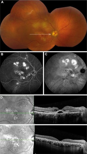
Table 2 Distribution of each criterion of severity among the severe cases of chronic central serous chorioretinopathy
Table 3 Characteristics at first presentation and disease progression in severe cases of chronic central serous chorioretinopathy
Table 4 Pearson correlation coefficients between patient characteristics and final visual outcome
Table 5 Frequency of all applied treatments in eyes with severe chronic central serous chorioretinopathy