Figures & data
Table 1 Enrolled patient characteristics
Figure 1 Flow diagram of the study.
Abbreviation: PDT, photodynamic therapy.
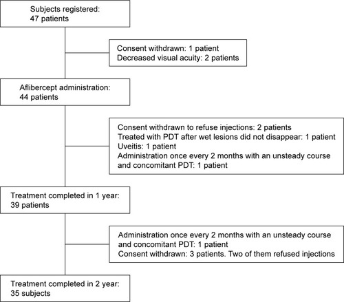
Table 2 Baseline characteristics of tAMD, type 1 PCV, and type 2 PCV
Figure 2 Indocyanine green angiogram of representative cases of type 1 and 2 PCV.
Notes: (A) Type 1 PCV at initial injection. (B) Type 1 PCV at 12 months. Polypoidal lesions are enlarged and number of polyps is increased. (C) Type 1 PCV at 24 months. Some polyps are enlarged and the lesions remain unchanged. (D) Type 2 PCV at initial injection. (E) Type 2 PCV at 12 months. Polypoidal lesions have disappeared. (F) Type 2 PCV at 24 months. No reappearance of polypoidal lesions.
Abbreviation: PCV, polypoidal choroidal vasculopathy.
Abbreviation: PCV, polypoidal choroidal vasculopathy.
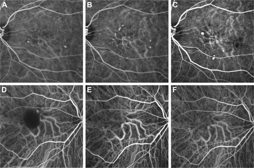
Figure 3 BCVA over 2 years of treatment (mean, SD).
Notes: Thirty-nine eyes with wet age-related macular degeneration and BCVA of ≥0.3 logMAR received three monthly intravitreal injections of aflibercept (IVA) followed by IVA every other month for up to 12 months and then with a modified treat-and-extend regimen from 12 to 24 months. Mean BCVA of 0.058 (20/22 Snellen equivalent) at 12 months significantly improved from initial mean BCVA of 0.097 (20/25 Snellen equivalent; paired t-test, P=0.03). Mean BCVA of 0.066 (20/23 Snellen equivalent) at 24 months was not significantly different from initial mean BCVA (paired t-test, P=0.09). *P<0.05.
Abbreviations: BCVA, best-corrected visual acuity; IVA, intravitreal aflibercept; logMAR, logarithm of minimum angle of resolution.
Abbreviations: BCVA, best-corrected visual acuity; IVA, intravitreal aflibercept; logMAR, logarithm of minimum angle of resolution.
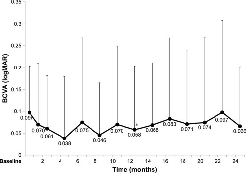
Figure 4 CMT over 2 years of treatment (mean, SD).
Notes: Thirty-nine eyes with wet age-related macular degeneration. Mean CMT significantly improved at 12 months (250 µm) and 24 months (240 µm) compared with baseline (320 µm) (paired t-test, P=0.002 and 0.0005, respectively). *P<0.05.
Abbreviation: CMT, central macular thickness.
Abbreviation: CMT, central macular thickness.
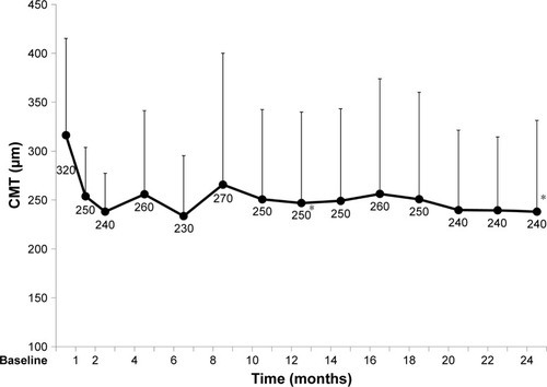
Figure 5 BCVA and CMT over 2 years of treatment in tAMD (tAMD [AMD] without PCV and retinal angiomatous proliferation) and PCV (mean, SD).
Notes: *P<0.05. (A) Solid line: 18 eyes with tAMD. Mean BCVA (0.070) at 12 months significantly improved from initial mean BCVA (0.13) (paired t-test, P=0.05). Mean BCVA (0.10) at 24 months is not significantly improved from initial mean BCVA (0.13) (paired t-test, P=0.2). Broken line: 21 eyes with PCV. Mean BCVA (0.048) at 12 months and (0.036) at 24 months are not significantly improved from initial mean BCVA (0.071) (paired t-test, P=0.2 and 0.2, respectively). (B) Solid line: mean CMT of tAMD patients tended to be improved at 12 months (260 µm) and 24 months (260 µm) than at baseline (320 µm) (paired t-test, P=0.08 and 0.07, respectively). Broken line: mean CMT of PCV patients significantly improved at 12 months (240 µm) and 24 months (220 µm) than at baseline (310 µm) (paired t-test, P=0.002 and <0.0001, respectively).
Abbreviations: AMD, age-related macular degeneration; BCVA, best-corrected visual acuity; CMT, central macular thickness; logMAR, logarithm of minimum angle of resolution; PCV, polypoidal choroidal vasculopathy; tAMD, typical age-related macular degeneration.
Abbreviations: AMD, age-related macular degeneration; BCVA, best-corrected visual acuity; CMT, central macular thickness; logMAR, logarithm of minimum angle of resolution; PCV, polypoidal choroidal vasculopathy; tAMD, typical age-related macular degeneration.
![Figure 5 BCVA and CMT over 2 years of treatment in tAMD (tAMD [AMD] without PCV and retinal angiomatous proliferation) and PCV (mean, SD).](/cms/asset/3216806e-e9a6-4408-a2df-5ce293d89367/doph_a_160961_f0005_c.jpg)
Figure 6 BCVA and CMT over 2 years of treatment in type 1 PCV and type 2 PCV (mean, SD).
Notes: *P<0.05. (A) Solid line: 12 eyes with type 1 PCV. Mean BCVA (0.059) at 12 months and (0.068) at 24 months are not significantly improved from initial mean BCVA (0.094) (paired t-test, P=0.4 and 0.07, respectively). Broken line: nine eyes with type 2 PCV. Mean BCVA (0.032) at 12 months and (−0.0062) at 24 months are not significantly improved from mean initial BCVA (0.040) (paired t-test, P=0.2 and 0.3, respectively). (B) Solid line: mean CMT of type 1 PCV tended to be improved at 12 months (230 µm) and 24 months (220 µm) than at baseline (320 µm) (paired t-test, P=0.09 and 0.03, respectively). Broken line: mean CMT of type 2 PCV improved at 12 months (250 µm) and 24 months (230 µm) than at baseline (300 µm) (paired t-test, P=0.04 and 0.02, respectively).
Abbreviations: BCVA, best-corrected visual acuity; CMT, central macular thickness; logMAR, logarithm of minimum angle of resolution; PCV, polypoidal choroidal vasculopathy.
Abbreviations: BCVA, best-corrected visual acuity; CMT, central macular thickness; logMAR, logarithm of minimum angle of resolution; PCV, polypoidal choroidal vasculopathy.
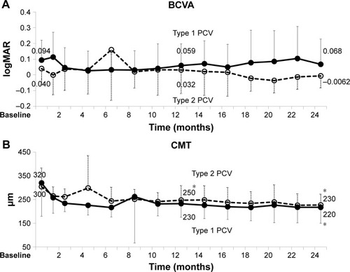
Table 3 Analysis of baseline characteristics and mean change of logMAR BCVA
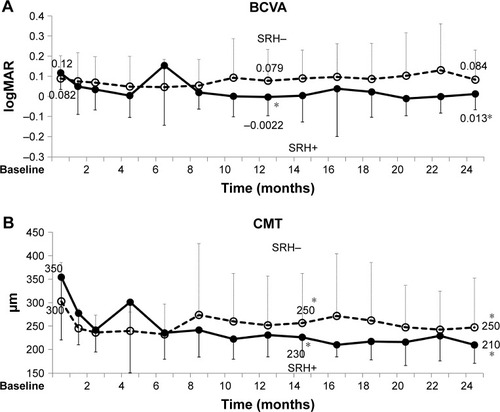
Figure 7 BCVA and CMT over 2 years of treatment in eyes with and without baseline SRH (mean, SD).
Notes: *P<0.05. (A) Solid line: 10 eyes with baseline SRH. Mean BCVA (−0.0022) at 12 months and (0.013) at 24 months are significantly improved than initial mean BCVA (0.12) (paired t-test, P=0.003 and 0.008, respectively). Broken line: 29 eyes without baseline SRH. Mean BCVA (0.079) at 12 months and (0.084) at 24 months are not significantly different from initial mean BCVA (0.082) (paired t-test, P=0.3 and 0.4, respectively). (B) Solid line: mean CMT of eyes with baseline SRH significantly improved at 12 months (230 µm) and at 24 months (210 µm) than at baseline (350 µm) (paired t-test, P=0.02 and 0.009, respectively). Broken line: mean CMT of eyes without baseline SRH significantly improved at 12 months (250 µm) and at 24 months (250 µm) than at baseline (300 µm) (paired t-test, P=0.03 and 0.01, respectively). Change of logMAR BCVA at 12 months is −0.12 with SRH and −0.011 without SRH (P=0.02). The change of logMAR BCVA at 24 months is −0.010 with SRH and −0.0056 without SRH (P=0.06).
Abbreviations: BCVA, best-corrected visual acuity; CMT, central macular thickness; logMAR, logarithm of minimum angle of resolution; PCV, polypoidal choroidal vasculopathy; SRH, subretinal hemorrhage.
Abbreviations: BCVA, best-corrected visual acuity; CMT, central macular thickness; logMAR, logarithm of minimum angle of resolution; PCV, polypoidal choroidal vasculopathy; SRH, subretinal hemorrhage.