Figures & data
Figure 1 Difference between uncorrected distance visual acuity vs corrected distance visual acuity (Snellen lines).
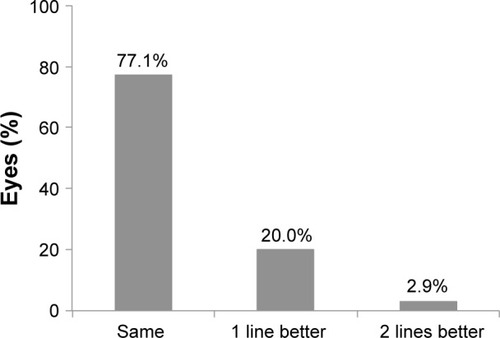
Figure 2 CDVA. Cumulative Snellen visual acuity (20/× or better). Twenty-four distance eyes.
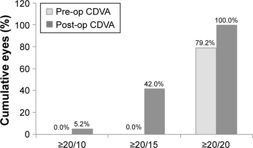
Figure 3 CDVA. Cumulative Snellen visual acuity (20/× or better). Eleven monovision eyes.
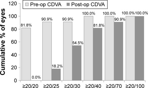
Figure 4 Sample case 1: 24-year-old male with 3 months post-LASIK follow-up data. Pre-op manifest: OD: +1.75 D, −0.50 × 123; OS: +5.25 D, −1.75 × 60. Measured treatment: OD: Contoura: plano, −0.59 × 164; WFO: +1.75; OS: Contoura: plano, −1.17 × 50; WFO: +5.00. Postop vision at 3 months: OD: 20/10- with refraction of plano; OS: 20/20 with refraction of plano, −0.75 × 15 (BCVA = 20/15). (A) is the OD (right) eye, (B) is the OS (left eye).
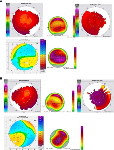
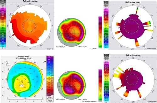
Figure 5 Sample case 2: 54-year-old female 5 months’ post-LASIK follow-up data. Pre-op manifest: OD: +2.25 D, −1.25 × 88; OS: +1.75 D, −0.75 × 80. Measured treatment: OD: Contoura: plano, −0.28 × 136; WFO: +3.25 (goal = −1.50 for monovision); OS: Contoura: plano, −0.20 × 7; WFO: +1.25. Post-op vision 5 months: OD: 20/30 with a refraction of −1.50 and J1 reading; OS: 20/15 with refraction of plano. (A) is OD and (B) is OS. Each figure shows pre-operative topography/pre-operative HOAs, postoperative topography/pre-operative Pentacam anterior elevation/Pre-operative anterior elevation treatment in Contoura surgical planning.
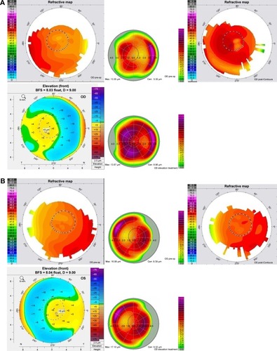
Figure 6 Sample case 3: 41-year-old male with 6 months’ post-LASIK follow-up data. Manifest refraction: OD: +6.00 D, −0.75 × 88; OS: +5.50 D, −0.50 × 92. Measured correction: OD: Contoura: plano, –0.38 × 73; WFO: +6.00; OS: Contoura: plano, –0.14 × 164; WFO: +5.50. Enhancement at 3 months treated via WFO: OD: −1.50 D, −0.50 × 30; OS: −2.00 D, −0.50 × 120. Postop vision at 6 months: OD: 20/20 with refraction of −0.50; OS: 20/15 with refraction of plano.