Figures & data
Figure 1 T2 MRI images of a patient with swollen lacrimal glands. The MRI images were obtained at the first visit to our hospital. The MRI findings indicate extreme swelling of both lacrimal glands.
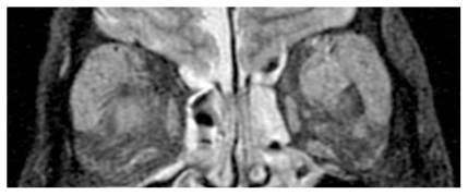
Figure 2 Histopathological findings in a specimen of the right lacrimal gland. H&E staining at low magnification (A × 100) and high magnification (B × 200) showing infiltration of plasma cells surrounding the mantle zones and a pattern of concentric deposits with lymphoid cells. These are pathognomic of Castleman disease. The infiltrated cells surrounding the mantle zones were CD138 (C × 100), Igkappa (E × 100), Iglambda (F × 100), and CD79a positive (D × 100). However, compared to plasma cell type of Castleman disease, fibrosis in the interstitial tissue was relatively greater. Compared to hyaline-vascular type of Castleman disease, vascularization in the intrafollicular area was relatively poor. The development of mantle zones and the pattern of concentric deposits with infiltrating cells are not of typical of Castleman disease. We finally decided these tissues resemble reactive lesions rather than Castleman disease.
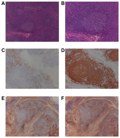
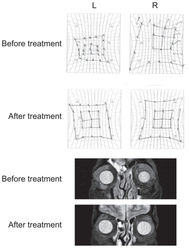
Figure 3 HESS charts and MRI findings before and after oral steroid administration. Both HESS charts and MRI findings were significantly improved after oral steroid administration. Six months after the treatment, no recurrence was observed. The effect of the oral steroid treatment was consistent with the histopathological findings of the specimen from the lacrimal glands.