Figures & data
Figure 1 Shows the intraoperative image of the technique. The ILM flap is stained with brilliant blue dye and then being raised and inverted into the MH using ILM peeling forceps.
Abbreviation: ILM, internal limiting membrane; MH, macular hole.
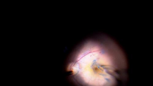
Figure 2 Intraoperative SD OCT image of inverted ILM flap being manipulated into the MH. The ILM flap segments are seen clearly on the OCT scan.
Abbreviations: ILM, internal limiting membrane; MH, macular hole; OCT, optical coherence tomography.
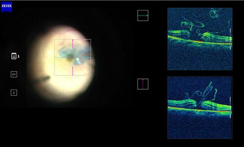
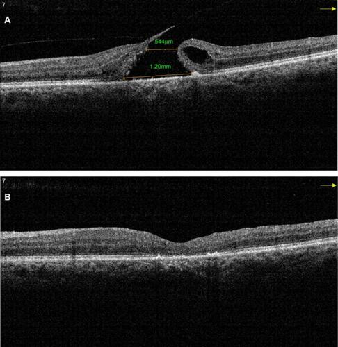
Figure 3 (A) Pre-operative SD OCT of a large MH with a horizontal linear width of 544 μm and base diameter of 1,200 μm. (B) Post-operative SD OCT of the same patient shows a U-type successful closure after vitrectomy with the inverted ILM flap technique.
Abbreviations: SD OCT, sSpectral domain optical coherence tomography; MH, mMacular hole.