Figures & data
Figure 1 Representative facial photos of two cases with IOI who presented with eyelid erythema, eyelid swelling and conjunctival hyperemia.
Notes: (A) A 73-year-old female and (B) a 51-year-old male. They were initially misdiagnosed as orbital cellulitis, however, antibiotics were ineffective. Their symptoms were rapidly resolved by systemic steroids, thus suggesting that they were suffering from IOI and not from orbital cellulitis.
Abbreviation: IOI, idiopathic orbital inflammation.
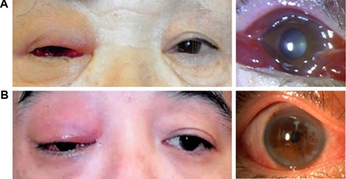
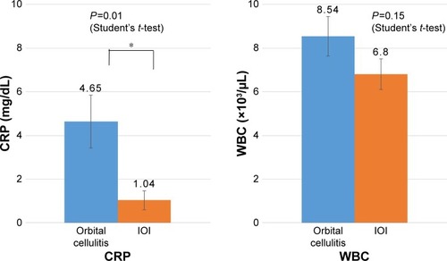
Figure 2 Levels of CRP and WBC count in the patients with orbital cellulitis and IOI.
Notes: The CRP levels were significantly higher in orbital cellulitis than in IOI, while the WBC levels were similar. *P<0.05.
Abbreviations: CRP, C-reactive protein; IOI, idiopathic orbital inflammation; WBC, white blood cell.
Abbreviations: CRP, C-reactive protein; IOI, idiopathic orbital inflammation; WBC, white blood cell.