Figures & data
Table 1 Demographics of the study population
Table 2 Mean MRSE and Km values for postoperative time points
Figure 1 Change in MRSE between postoperative time points for the entire patient cohort.
Abbreviations: MRSE, manifest refractive spherical equivalent; preop, preoperative.
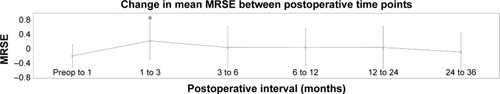
Figure 2 Change in MRSE with respect to baseline MRSE for all postoperative follow-up time points.
Abbreviation: MRSE, manifest refractive spherical equivalent.
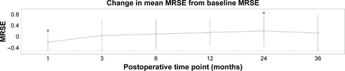
Figure 3 Change in Km between postoperative time points for the entire patient cohort.
Abbreviations: Km, mean keratometry; preop, preoperative.
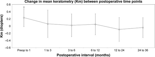
Figure 4 Change in Km with respect to baseline Km for all postoperative follow-up time points.
Abbreviation: Km, mean keratometry.
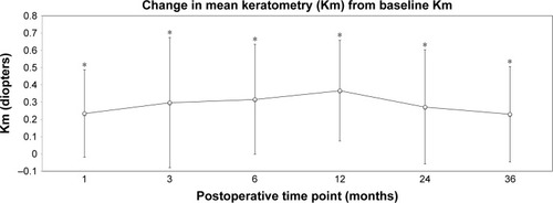
Figure 5 Demonstration of topographic pattern of annular steepening with central flattening occurring over the 36-month follow-up period with a corresponding hyperopic shift in four patients (#1–4).
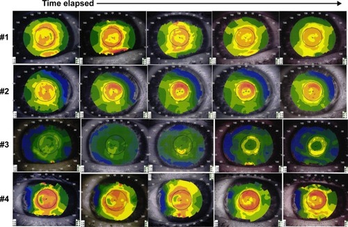
Figure 6 Longitudinal corneal topographic changes associated with MRSE changes in a single patient. Pronounced correlation of topographic “ring” disappearance and myopic regression in late postoperative interval.
Abbreviations: MRSE, manifest refractive spherical equivalent; postop, postoperative; preop, preoperative.
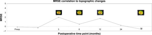
Figure 7 (A) Central steepening and annular flattening topography at 36 months in a patient with significant myopic shift. (B) Central flattening and annular steepening topography at 36 months in a patient with significant hyperopic shift.
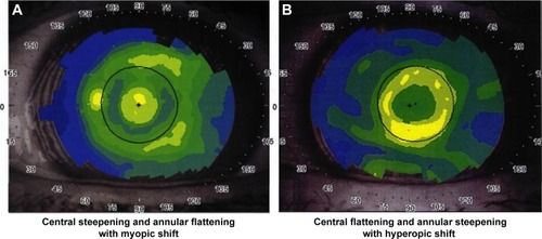
Table 3 SIA between postoperative time points
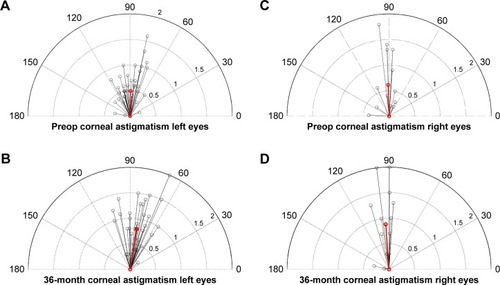
Figure 8 Depictions of corneal astigmatisms and axis for left and right eyes in vector format.
Abbreviation: preop, preoperative.