Figures & data
Figure 1 Example of a case of POAG with inferior retinal nerve fiber layer defect characterized with a superior arcuate defect on VF.
Abbreviations: POAG, primary open-angle glaucoma; VF, visual field.
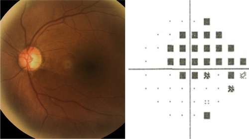
Table 1 Demographic and clinical characteristics by presence of POAG
Figure 2 Curve of intraocular pressure according to POAG diagnosis.
Abbreviation: POAG, primary open-angle glaucoma.
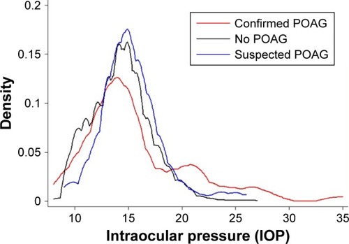
Table 2 Distribution and relationship between POAG diagnoses according to the blood pressure level
Table 3 Distribution and relationship between POAG diagnosis according to the OPP
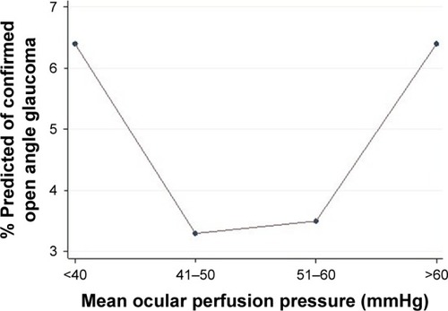
Figure 3 Relationship between OPP and confirmed POAG.
Abbreviations: OPP, ocular perfusion pressure; POAG, primary open-angle glaucoma.