Figures & data
Figure 1 Steps of our 27-gauge needle-assisted externalization and haptic securing technique.
Notes: (A) Labeling 2mm from limbus-180 degree apart. (B) 27G needle introduction aiming mid vitreous. (C) Externalized via main tunnel-guided by visco-canula. (D) Loading the IOL haptic into the 27G needle. (E) Externalizing the haptic. (F–H) Same procedure repeated on the other side. (I) Tunnel creation for using 27G needle. (J–L) Docking the haptic into the scleral tunnel.
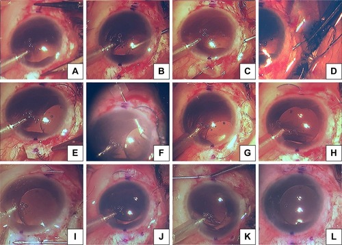
Figure 2 3D – steps of our 27-gauge needle-assisted externalization and haptic securing technique.
Notes: (A) Entry of the 27G needle aiming mid vitreous. (B) Visco-Canula used to engage the tip of the 27G needle and externalisation. (C) Loading of the haptic into the lumen of the syringe. (D) Externalization of the haptic-secured by stopper. (E) Same procedure repeated on the other side. (F) Tunnel creation for the haptic docking using 27G needle. (G) Docking the haptic.
Abbreviation: 3D, three dimensional.
Abbreviation: 3D, three dimensional.
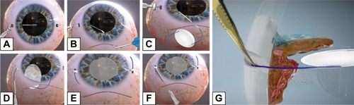
Figure 3 Method of loading the “stopper” to the 27-gauge needle which prevents the haptic from slipping back into the vitreous cavity after externalization.
Notes: (A) Silicon sleeve – which later acts as stopper being loaded to the bent 27-gauge needle. (B) Final position of the stopper.
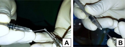
Table 1 Baseline, indications and postoperative characteristics of scleral-fixated IOL
Table 2 Special cases: their baseline and postoperative characteristics
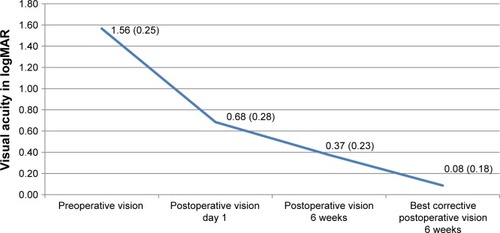
Figure 4 The pattern of preoperative, postoperative, and 6th week postoperative change in visual acuity.