Figures & data
Table 1 Shows the main refractive outcomes over 18 months of follow-up
Table 2 Intraoperative and postoperative complications of femtosecond-assisted intracorneal stromal ring
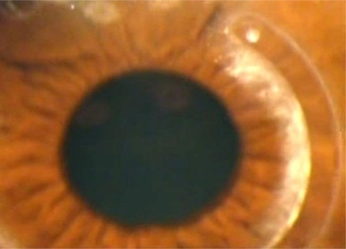
Figure 1 (A) Scheimpflug tomography by Sirius CSO showing decentered ring. (B) Scheimpflug image of the cornea showing decentered rings near toward the pupil. (C) Cornea view of the eye showing the Keraring crossing the pupil.
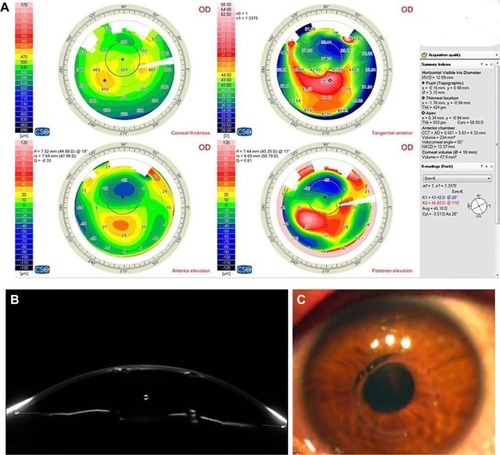
Figure 2 (A) Signs of infection in the presence of Kerarings. (B) Explantation of Keraring. (C) Postexplantation corneal scarring.
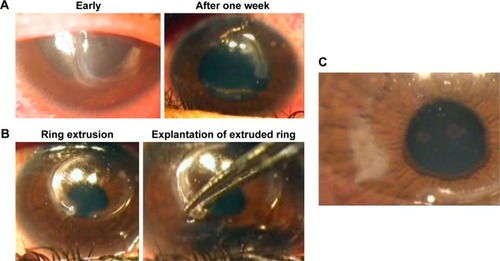
Figure 3 Crystalline deposits around the Keraring.