Figures & data
Figure 1 Marginal tear strip as seen by AS-SD-OCT.
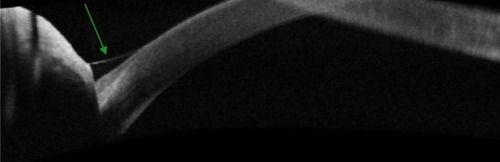
Figure 2 Tear meniscus parameters as measured by AS-SD-OCT.
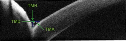
Table 1 Mean, SD and range of TMH in the different LASIK techniques at different times of follow-up
Table 2 Mean, SD and range of TMD in the different LASIK techniques at different times of follow-up
Table 3 Mean, SD and range of TMA in the different LASIK techniques at different times of follow-up
Table 4 P-value in pairwise comparison of the TMH between the three LASIK techniques at different times of follow-up
Figure 3 In all techniques, TMH dropped after 1 week to the lowest level.
Abbreviations: TMH, tear meniscus height; FS-SMILE, femtosecond small incision lenticule extraction; FS-LASIK, femtosecond laser in situ keratomileusis; LASIK, laser in situ keratomileusis; CI, confidence interval.
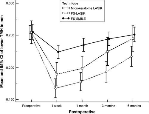
Table 5 P-value in pairwise comparison of the TMD between the three LASIK techniques at different times of follow-up
Figure 4 In all techniques, TMD dropped after 1 week to the lowest level.
Abbreviations: TMD, tear meniscus depth; LASIK, laser in situ keratomileusis; FS-SMILE, femtosecond small incision lenticule extraction; FS-LASIK, femtosecond laser in situ keratomileusis; CI, confidence interval.
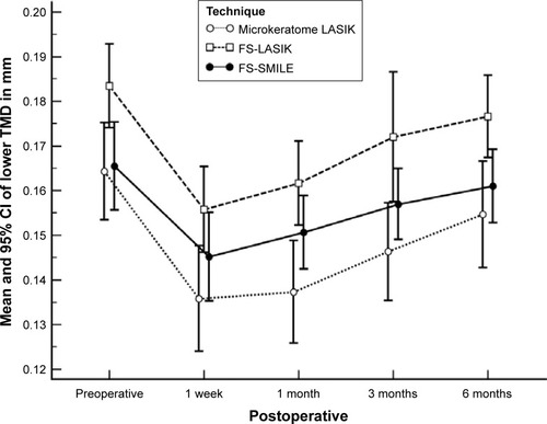
Table 6 P-value in pairwise comparison of TMA between the three LASIK techniques at different times of follow-up
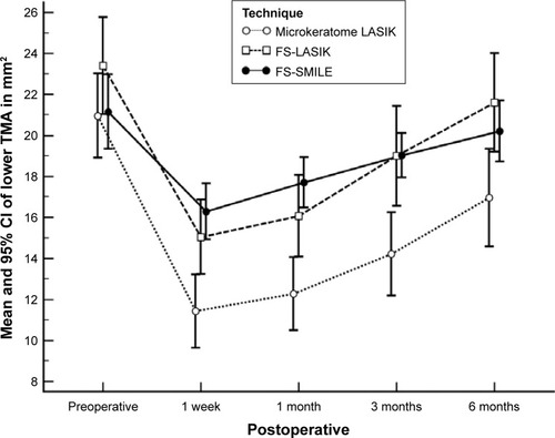
Figure 5 In all techniques, TMA dropped after 1 week to the lowest level.
Abbreviations: TMA, tear meniscus area; FS-SMILE, femtosecond small incision lenticule extraction; FS-LASIK, femtosecond laser in situ keratomileusis; LASIK, laser in situ keratomileusis; CI, confidence interval.