Figures & data
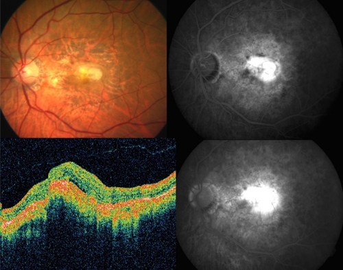
Figure 1 Case 1: Color fundus photograph of the right eye (upper left) shows a grayish-brown subfoveal lesion suggestive of choroidal neovascular membrane. Early hyperfluoresence with intense late staining on fundus fluorescein angiography confirms the presence of predominantly classic choroidal neovascularization (upper right and lower right). Optical coherence tomography scan reveals the presence of a high reflective, spindle-shaped lesion abutting the fovea with increased overlying retinal thickness and intraretinal cystoid spaces (lower left).
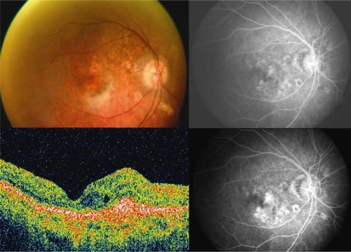
Figure 2 Sequential comparison of optical coherence tomography scans over time for the three study eyes. Column 1 denotes Case 1: At presentation (left, top), two months after initial treatment (left, middle), and at last follow-up (left, bottom). Column 2 denotes Case 2: At presentation (central, top), four weeks after initial treatment (central, middle), and at last follow-up (central, bottom). Column 3 denotes Case 3: At presentation (right, top), four weeks after initial treatment (right, middle), and at last follow-up (right, bottom). Cases 1 and 3 reveal progressive retinal thinning at the macula, while Case 2 reveals retinal thinning with evolution of full- thickness macular hole.
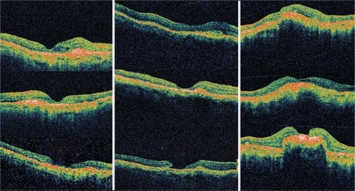
Figure 3 Case 2: Color fundus photograph of the right eye (upper left) shows a yellowish-gray subfoveal lesion suggestive of choroidal neovascularization with specks of fresh subretinal hemorrhage. Early hyperfluoresence with intense late staining on fundus fluorescein angiography confirms the presence of predominantly classic choroidal neovascularization (upper right and lower right); blocked fluorescence corresponds to subretinal hemorrhage. Optical coherence tomography scan reveals the presence of a high reflective, spindle-shaped lesion in the subretinal area abutting the fovea with increased overlying retinal thickness (lower left).
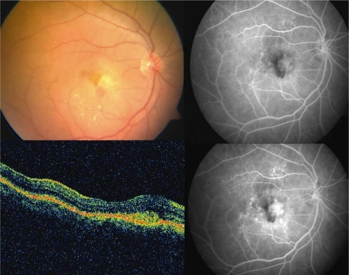
Figure 4 Case 3: Color fundus photograph of the left eye (upper left) shows a partially regressed choroidal neovascularization. Early hyperfluoresence with intense late staining on fundus fluorescein angiography confirms the presence of choroidal neovascularization (upper right and lower right). Optical coherence tomography scan reveals the presence of subfoveal fluid and cystoid macular edema associated with the choroidal neovascularization (lower left) in the left eye.