
Figures & data
Table 1 8K UHD CMOS imaging sensor specifications
Figure 1 Our new 8K UHD camera mounted on a surgical microscope with a relay lens adapter.
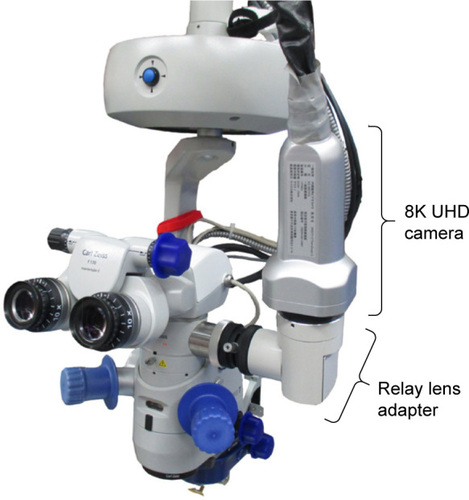
Figure 2 8K UHD microscopic camera for ophthalmic surgery in the operating room site.
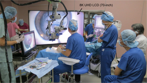
Figure 3 The comparison of surgical field images of pig cadaver eye in cataract surgery (phacoemulsification). (A) Image obtained with the prototype 8K microscopic camera and (B) image obtained with the new 8K UHD microscopic camera.
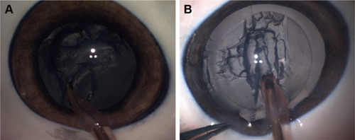
Figure 4 The comparison of surgical field images of pig cadaver eye in glaucoma surgery. (A) Images obtained with the prototype 8K microscopic camera and (B) images obtained with the new 8K UHD microscopic camera through a gonioscope.
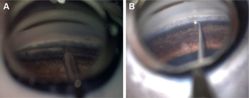
Figure 5 Comparison of surgical field images of pig cadaver eye in vitreoretinal surgery. (A) Images obtained with the prototype 8K microscopic camera and (B) images obtained with the new 8K UHD microscopic camera. Retinal capillary vessels were illuminated by a chandelier endo-illumination fiber.
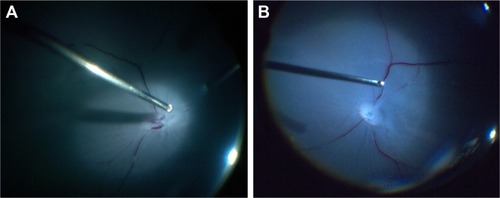
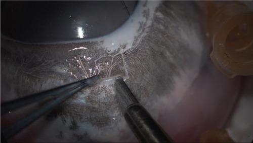
Figure 6 8K UHD image in creating a sclera flap of pig cadaver eye.
Video S1 Detail of – with HD resolution down-converted from an original 8K UHD video.
Abbreviations: HD, high-definition; UHD, ultra-high-definition.