Figures & data
Figure 1 Anterior segment optical coherence tomography imaging. The picture shows a persisting irido-corneal contact despite central deepening of the anterior chamber during corneal indentation (synechial closure).
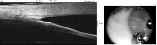
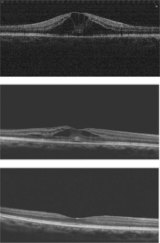
Figure 2 Spectral domain OCT macular images showing the evolution of cystoid macular edema, gradually improving and completely recovering after four weeks (lower picture) of medical treatment.