Figures & data
Table 1 Intraclass correlation coefficients of central corneal thickness measurements in normal subjects
Table 2 Repeated-measure analysis of variance with Bonferroni post hoc comparison, LOA, and ICC
Figure 1 Comparisons of central corneal thickness (CCT) measurement among the four devices.
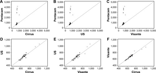
Figure 2 Bland–Altman plots showing agreement of central corneal thickness (CCT) measurements among the four devices.
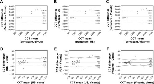
Figure 3 Comparison of interobserver and intraobserver repeatability between two central corneal thickness (CCT) measurements from each of the four devices. Notes: Correlations between the two examinations by one examiner of CCT measurement by (A) the Pentacam, (B) Visante optical coherence tomography (OCT), (C) Cirrus OCT, and (D) ultrasound (US) pachymetry. Correlations between the two examiners of CCT measurement by (E) the Pentacam, (F) Visante OCT, (G) Cirrus OCT, and (H) US. A1, first measurement by examiner 1; A2, second measurement by examiner 1; B1, first measurement by examiner 2.
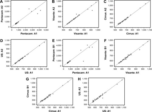
Table 3 Central corneal thickness measurements from each device compared by corneal thickness
Table 4 Mean difference ± SD, LOA, ICCs, and correlations between the pairs of methods
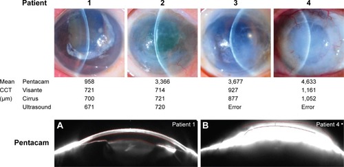
Figure 4 Central corneal thickness measurements in each grade of corneal edema and anterior-segment photos obtained using the Pentacam with inexact anterior and posterior corneal alignment.