Figures & data
Figure 1 One-handed coaxial phacoemulsification technique.
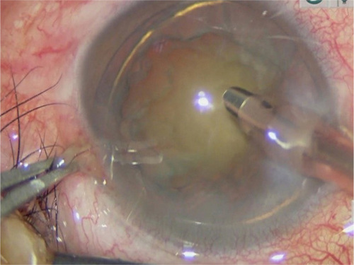
Figure 2 Two-handed coaxial phacoemulsification technique.
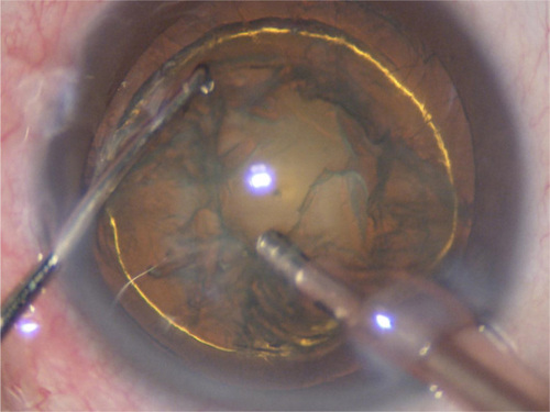
Table 1 Patient demographics and baseline parameters
Table 2 Intraoperative parameters of the 2 groups
Table 3 Visual acuity and SIA
Table 4 CV, CCT and corneal endothelial cell counts/size
Figure 3 Change in corneal volume over time.
Abbreviations: w, week; m, months; pre, preoperative.
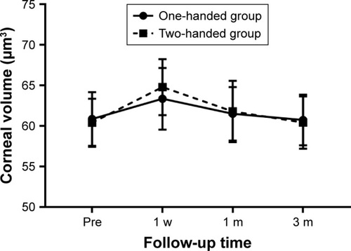
Figure 4 Change in central corneal thickness over time.
Abbreviations: w, week; m, months; pre, preoperative.
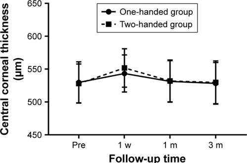
Figure 5 Change in endothelium corneal density over time.
Abbreviations: w, week; m, months; pre, preoperative.
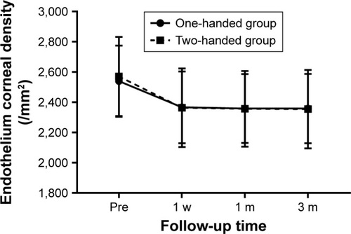
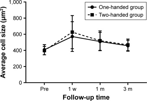
Figure 6 Change in average cell size over time.
Abbreviations: w, week; m, months; pre, preoperative.