
Figures & data
Figure 1 Time-domain optical coherence tomography (Cirrus™, Carl Zeiss Meditec) findings from Case 1.
Notes: OD and OS imaging at initial presentation with sub-foveal nodules present. Top images are a cross section of the fovea in the nasal-temporal orientation, bottom images are a cross section in the inferior-superior orientation.
Abbreviations: N, nasal; T, temporal; S, superior; I, inferior.
Abbreviations: N, nasal; T, temporal; S, superior; I, inferior.
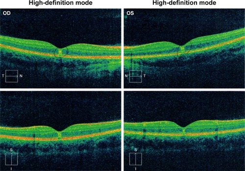
Figure 2 Time-domain optical coherence tomography (Cirrus™, Carl Zeiss Meditec) findings from Case 1.
Notes: OD and OS imaging from 2-month follow up, with no sub-foveal nodules noted. Top images are a cross section of the fovea in the nasal-temporal orientation, bottom images are a cross section in the inferior-superior orientation.
Abbreviations: N, nasal; T, temporal; S, superior; I, inferior.
Abbreviations: N, nasal; T, temporal; S, superior; I, inferior.
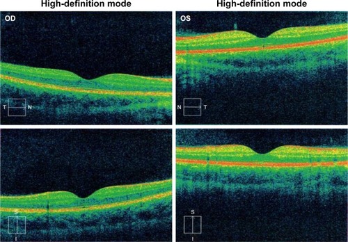
Figure 3 Time-domain OCT (Cirrus™, Carl Zeiss Meditec) from Case 2.
Notes: Inner segment/outer segment junction abnormalities in OD and OS consistent with solar retinopathy. Image on the left shows the left eye, image on the right shows the right eye.
