
Figures & data
Figure 1 Surgical manipulations in Case 1.
Notes: PEA was performed by widening the pupil with iris retractors. After confirming that the Zinn’s zonule dialysis ranged from the 2 to 8 o’clock position, we removed the remaining lens nucleus by visco-extraction with a Simcoe aspiration needle. The procedure performed after the aspiration was as follows. (A) Viscoelastic material was packed into the collapsed capsular bag in the dialysis portion to re-inflate it to nearly normal. (B) A scleral flap was created at the 5 o’clock position. (C) A 27G needle was inserted under the scleral flap. A Pair-Pak® needle was inserted from the opposite side in order to pierce through the equatorial segment of the capsular bag. (D) The Pair-Pak needle was pushed from the interior of the eye to outside the sclera. (E) The preceding IOL loop was bound to the thread of the Pair-Pak needle that was inserted into the interior of the eye. (F) After inserting the posterior loop into the capsular bag, the thread of the preceding loop was sutured inside the scleral flap.
Abbreviations: IOL, intraocular lens; PEA, phacoemulsification and aspiration.
Abbreviations: IOL, intraocular lens; PEA, phacoemulsification and aspiration.
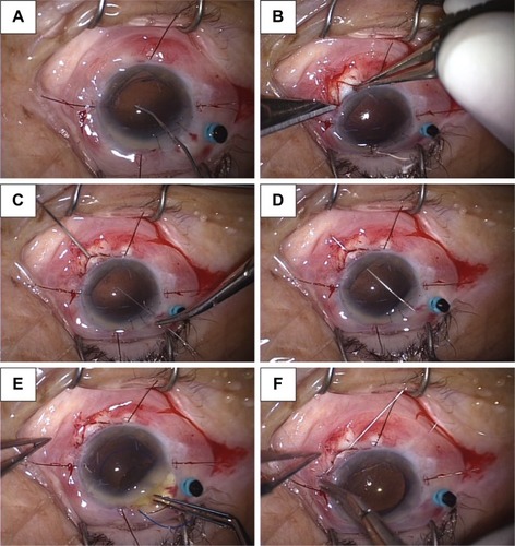
Figure 2 (A) Zinn’s zonule dialysis is observed in the right side. (B) The straight needle of Pair-Pak passes through the path of the 27 G needle, it pierces the equatoria segment. (C) At the end of the surgery, the unilateral IOL loop is fixed with a 10-0 polypropylene thread from the scleral flap.
Abbreviation: IOL, intraocular lens.
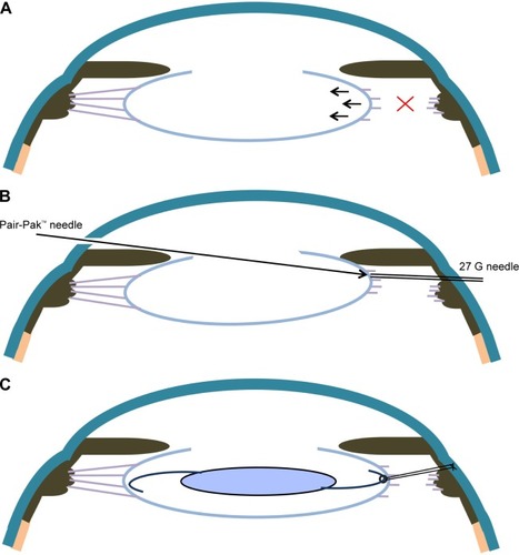
Figure 3 Case 1: Preoperative anterior segment of the right eye.
Notes: (A) A cataract and shallow anterior chamber were observed. (B) Moderate mydriasis was observed. (C), (D) Anterior segment of the right eye at 1 week after the operation. The anterior chamber was deep and did not exhibit any IOL inclination or deflection. The pupil was nearly circular.
Abbreviation: IOL, intraocular lens.
Abbreviation: IOL, intraocular lens.
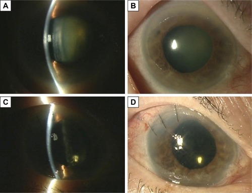
Table 1 Preoperative, intraoperative, and postoperative states in cases treated with this procedure