
Figures & data
Figure 1 Optical coherence tomography of a tractional lamellar macular hole, characterized by epiretinal membrane with surface wrinkling, sharp intraretinal split, and “schisis-like” appearance.
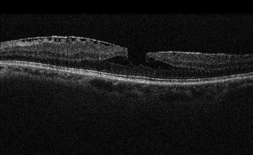
Figure 2 Degenerative lamellar macular hole identified by round-edged intraretinal cavitation, “foveal bump”, and epiretinal proliferation of medium reflectivity.
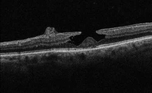
Table 1 Studies on natural history of lamellar macular holes
Table 2 Studies describing surgical outcomes of vitrectomy for lamellar macular holes