Figures & data
Figure 1 The Cirrus HD-OCT macular cube 200×200 protocol provides regional GCIPL thickness in six wedge-shaped sectors.
Abbreviation: GCIPL, ganglion cell-inner plexiform layer.
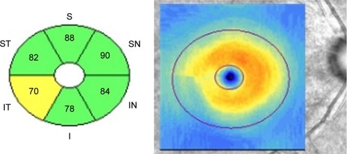
Table 1 Demographic and clinical characteristics of the study population
Table 2 Comparison of cpRNFL thickness, GCIPL thickness and asymmetry measurements, and ONH parameters between two groups
Table 3 AUROC and pAUROC values for cpRNFL thickness, GCIPL thickness, GCIPL asymmetry measurements, and ONH parameters
Table 4 P-values for pairwise comparison of AUROC values and pAUROC values between the best measure of each GCIPL asymmetry analysis and cpRNFL, GCIPL, and ONH parameters
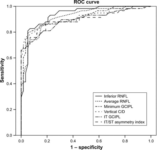
Figure 2 AUROC values for OCT parameters to discriminate early glaucoma with paracentral scotoma from normal controls.
Note: The inferior RNFL thickness had the largest AUROC value (0.934), followed by average RNFL thickness (0.914), minimum GCIPL thickness (0.911), vertical C/D (0.910), IT GCIPL thickness (0.896), and IT/ST asymmetry index (0.894).
Abbreviations: AUROC, area under the receiver operating characteristic curve; C/D, cup-to-disc ratio; GCIPL, ganglion cell-inner plexiform layer; IT, inferotemporal; OCT, optical coherence tomography; RNFL, retinal nerve fiber layer; ROC, receiver operating characteristic; ST, superotemporal.
Abbreviations: AUROC, area under the receiver operating characteristic curve; C/D, cup-to-disc ratio; GCIPL, ganglion cell-inner plexiform layer; IT, inferotemporal; OCT, optical coherence tomography; RNFL, retinal nerve fiber layer; ROC, receiver operating characteristic; ST, superotemporal.