
Figures & data
Figure 1 Vernal keratoconjunctivitis (VKC): the tarsal form with giant papillae (left panel) and limbal form with Horner-Trantas dots (right panel, black and white arrows).
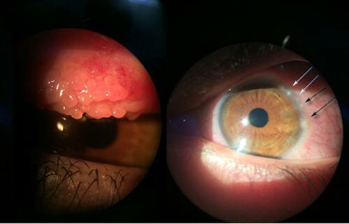
Figure 2 Vernal keratoconjunctivitis (VKC) corneal alterations as observed with confocal microscopy. (A) Larger and hyperreflective epithelial cells; (B) tortuous superficial nervous plexus; and (C–D) higher concentration of inflammatory cells and activated keratocytes.
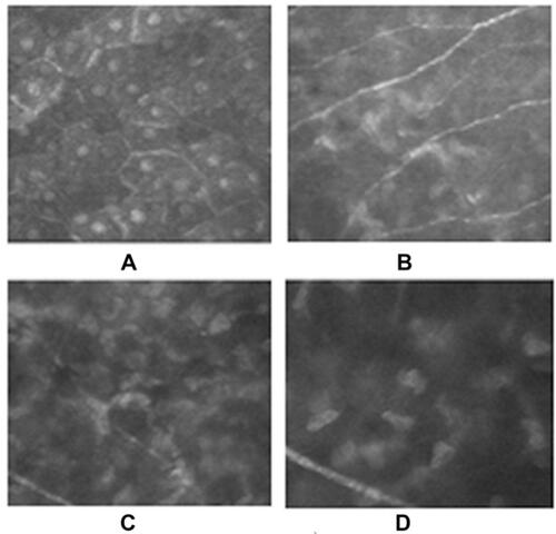
Table 1 Most common concentration of CyA eye drops formulations and countries of distribution
Table 2 Summary of 0.1% (1 mg/mL) concentration CyA studies in VKC patients