Figures & data
Table 1 Baseline patient characteristics
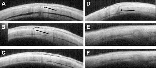
Figure 1 Time course of logMAR BCVA.
Notes: The mean logMAR BCVA at baseline and 1, 3, and 6 months postoperatively was 0.107±0.231, 0.022±0.130, 0.003±0.105, and 0.0001±0.102, respectively, in the straight group, and 0.079±0.178, 0.112±0.288, 0.095±0.222, and 0.090±0.225, respectively, in the angled group. No difference in the logMAR BCVA between the two groups was significant during follow-up (P>0.05, respectively).
Abbreviations: BCVA, best-corrected visual acuity; logMAR, logarithm of minimal angle of resolution; ns, not significant.
Abbreviations: BCVA, best-corrected visual acuity; logMAR, logarithm of minimal angle of resolution; ns, not significant.
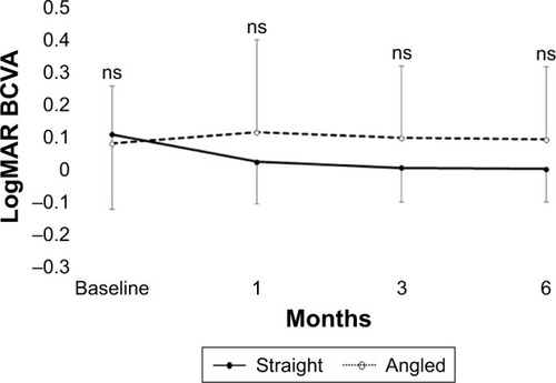
Figure 2 Time course of changes in IOP.
Notes: The mean IOP at baseline was 14.9±3.6 mmHg in the straight group and 14.8±3.6 mmHg in the angled group. At 1, 2, 3, and 10 days postoperatively, IOP was 11.5±5.9, 12.1±6.3, 11.9±4.2, and 15.0±3.6 mmHg, respectively, in the straight group, and 13.4±7.4, 12.9±7.0, 12.6±3.9, and 14.2±4.3 mmHg, respectively, in the angled group. No difference in IOP between the two groups was significant during follow-up. However, the mean IOP in the early postoperative period was significantly lower than that at baseline in the straight group (day 1, P=0.010; day 2, P=0.002; day 3, P=0.030). *P<0.05 vs baseline.
Abbreviations: IOP, intraocular pressure; ns, not significant.
Abbreviations: IOP, intraocular pressure; ns, not significant.
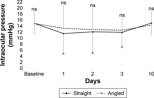
Table 2 Rate of wound closure of each port
Figure 3 Anterior segment OCT images of sclerotomies.
Notes: (A–C) OCT images of sclerotomies of patients with straight incisions at 1, 3, and 10 days postoperatively. (A and B) Significant gaps (arrows) in the sclera are visible as hyporeflective areas 1 and 3 days postoperatively. (C) However, the hyporeflective area is not observed in the sclera 10 days postoperatively. (D–F) OCT images of sclerotomies of patients with angled incisions 1, 3, and 10 days postoperatively. (D) A significant gap (arrow) in the sclera is visible as a hyporeflective area 1 day postoperatively. (E and F) However, no gap in the sclera is observed from 3 days postoperatively.
Abbreviation: OCT, optical coherence tomography.
Abbreviation: OCT, optical coherence tomography.