
Figures & data
Figure 1 Nerve supply of the cornea. The cornea is innervated by the ophthalmic branch of the trigeminal nerve (V1) and by sympathetic and parasympathetic autonomic nerves.
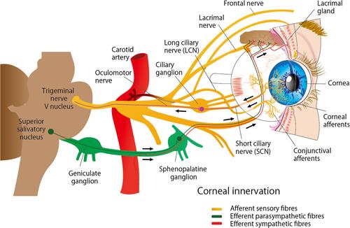
Figure 2 Corneal nerves distribution: Nerve fibers penetrate the corneal periphery at the limbus and approach toward the central cornea at the level of the anterior stroma while penetrating the Bowman’s membrane to create the sub-basal nerve plexus, at the level of the basal epithelial cells and basement membrane of the epithelium. Terminal branches from the sub-basal plexus pass anteriorly into the epithelial cell layers, terminating within or in between epithelial cells.
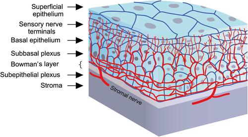
Figure 3 Stages of neurotrophic keratitis: Impaired cornea sensitivity and lack of trophic support trigger nonspecific epithelial irregularity and tear film changes (A, B). Stage 1- mild (C, D) is characterized by corneal punctate keratopathy due to focal epithelial damage and loss of tight junctions. It is associated with mild stromal edema with or without corneal neovascularization. Stage 2 – moderate (E, F) is distinguished by the presence of central persistent epithelial defect, surrounded by edematous, cloudy, and poorly adherent epithelium. Stage 3- severe (G, H) is characterized by stromal ulceration and thinning that may progress to melting and perforation (arrows).
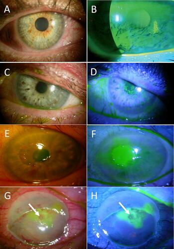
Table 1 Summary Of Clinical Studies Evaluating Safety And Efficacy Of Recombinant Human Nerve Growth Factor (rhngf) Eye Drops