Figures & data
Figure 1 Imaging of a donor cornea through a sealed and sterile case.
Abbreviation: HD-OCT, high definition optical coherence tomography.
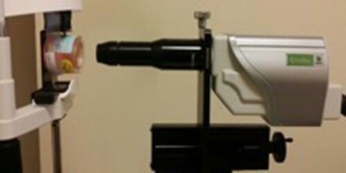
Figure 2 HD-OCT images from (A) donor graft; (B) control eye; and (C) Fuchs’ endothelial corneal dystrophy eye. Below each HD-OCT image is the result of segmentation showing the isolated En/DM complex demarcated with red arrows. Images are displayed with the zero delay at the bottom.
Abbreviations: HD-OCT, high definition optical coherence tomography; En/DM, endothelial/Descemet membrane.
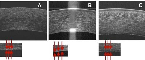
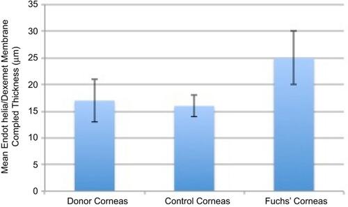
Figure 3 Mean En/DM complex thickness (μm) in grafts versus control eyes and eyes with Fuchs’ endothelial corneal dystrophy.
Abbreviation: En/DM, endothelial/Descemet membrane.