Figures & data
Figure 1 A) Anterior chamber image. The NewIris lens is positioned over the whole surface of the iris. Corneal incision to insert the lens is at twelve o’clock with a size of 2.8 mm; B) Iris lens diaphragm after explantation.
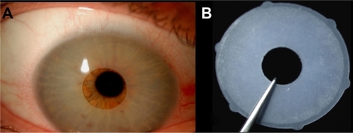
Figure 2 Endothelial cell count. The polymorphism and polymegathism are very evident in both eyes, especially the left one.
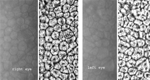
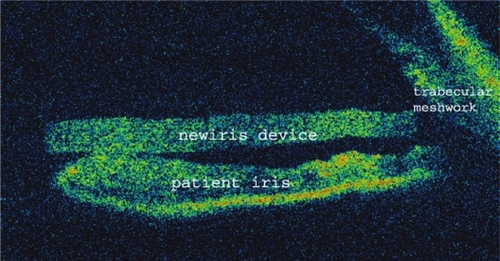
Figure 3 Anterior chamber optical coherence tomography. The lens is positioned over the iris and is in contact with all the 360° coneoescleral trabecular meshwork.