
Figures & data
Figure 1 Infrared images showing a typical pattern of “starry sky” fundus with numerous tiny glittering crystal deposits throughout the entire posterior pole of a patient with BCD (both eyes).
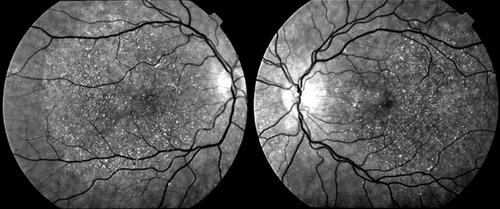
Figure 2 Color retinography of the fundus of both eyes of different patients showing different degrees of evolution of BCD disease, according to Yuzawa classification. (A) Numerous glistening crystalline dots and minimal chorioretinal damage restricted to the posterior pole of the retina, corresponding with stage 1 of Yuzawa. (B) Epithelial confluent atrophy of the posterior pole and few crystalline deposits, corresponding with stage 2 of Yuzawa. (C) Advanced choroidal sclerosis with marked chorioretinal atrophy and absence of deposits, corresponding with stage 2 of Yuzawa. Misdiagnosing with severe forms of choroideremia or retinitis pigmentosa is possible.
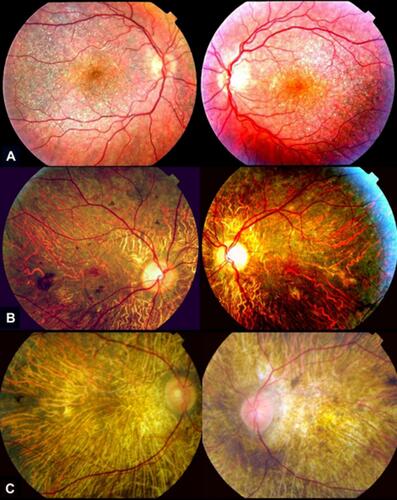
Table 1 Presence of corneal crystalline deposits in patients with clinical and genetic diagnosis of BCD confirmed by CYP4V2 gene analysis, according to the medical literature.
Figure 3 Slit-lamp biomicroscopic findings in several patients with BCD with corneal involvement. Crystalline-like corneal deposits (arrows) can be present in 360º degrees of the limbus area of both eyes.
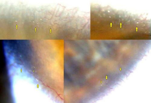
Figure 4 In vivo confocal microscopy image (400×400 μm) of a BCD patient (subepithelium area). The crystals (yellow arrows) were up to 24 μm in length and 16 μm in width and barely distinguishable from keratocytes nuclei (blue arrows).
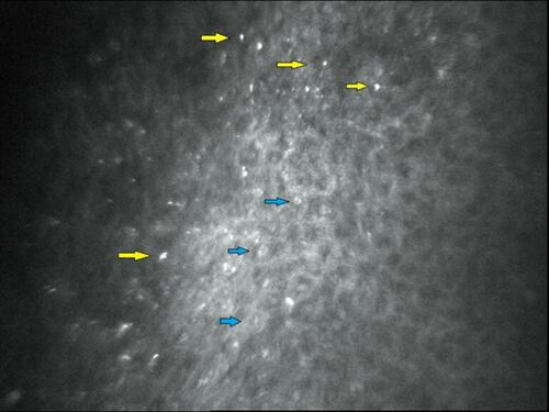
Figure 5 Fluorescein angiography in later stage on both eyes of different patients with genetic diagnosis of BCD. (A) Small-patched areas of atrophy of the choriocapillaris and RPE, the macular area remain relatively intact in an early stage of BCD. (B) Marked atrophy of the RPE with focal confluent areas of disappearance of the choriocapillaris. (C) Severe atrophy of the RPE with complete disappearance of the choriocapillaris.
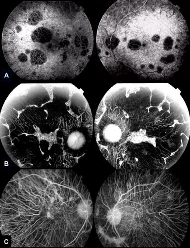
Figure 6 Spectral domain-optical coherence tomography (SD-OCT) image revealing both outer retinal tabulation (blue arrow) and crystalline macular deposits (yellow arrow) of a BCD individual.
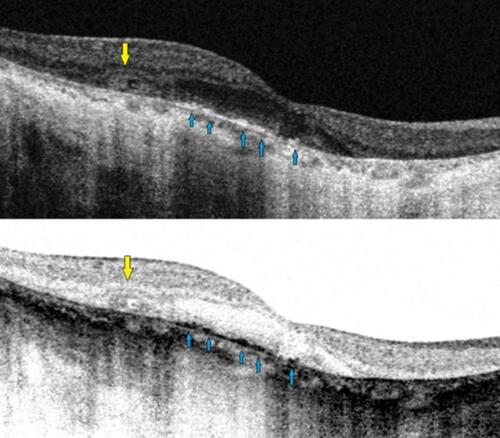
Table 2 Summary of the mutations identified in CYP4V2 gene in patients with molecular diagnosis of BCD, according to the medical literature.