Figures & data
Figure 1 Widefield color fundus photography from an affected CHM male patient. Widefield images show areas of chorioretinal atrophy mainly localised in the periphery and peripapillary regions (red arrows). Images of this patient courtesy of Dr. Andrea Scupola, MD.
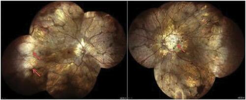
Figure 2 Multimodal imaging from an affected CHM male patient. Multicolor images (left) show areas of RPE atrophy in the macula. Blue fundus autofluorescence (middle left) and fluorescein angiography (middle right) images display the presence of RPE and choroidal atrophy, respectively. Structural OCT images (right) demonstrate a thin choroid and an RPE and outer retinal atrophy. Images of this patient courtesy of Prof. Eric Souied, MD, PhD.
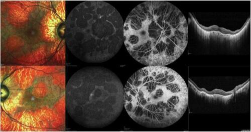
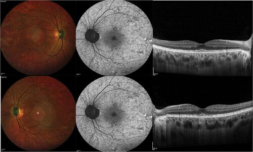
Figure 3 Multimodal imaging from a female CHM carrier. Multicolor images (left) show areas of RPE alteration and mottling in the macula. Blue fundus autofluorescence images (middle) show a characteristic pattern of speckled autofluorescence. Structural OCT images (right) demonstrate a normal appearance of the macular retina and choroid.