
Figures & data
Table 1 Demographics of 32 eyes from 28 patients who underwent DMEK (or triple DMEK)
Figure 1 Representative cases with surgery-induced iris abnormalities after DMEK. (A) Case 1: a 69-year-old man had bullous keratopathy after cytomegalovirus corneal endotheliitis in his right eye. This eye was pseudophakic. (B) Case 1, after solitary DMEK, mild ovalization of the pupil (arrow), likely due to posterior synechiae, was noted. (C) Case 2: a 73-year-old woman had laser iridotomy-related bullous keratopathy in her right eye. This eye was pseudophakic. (D) Case 2, after solitary DMEK, mild iris depigmentation (arrows) was detected near the temporal wound.
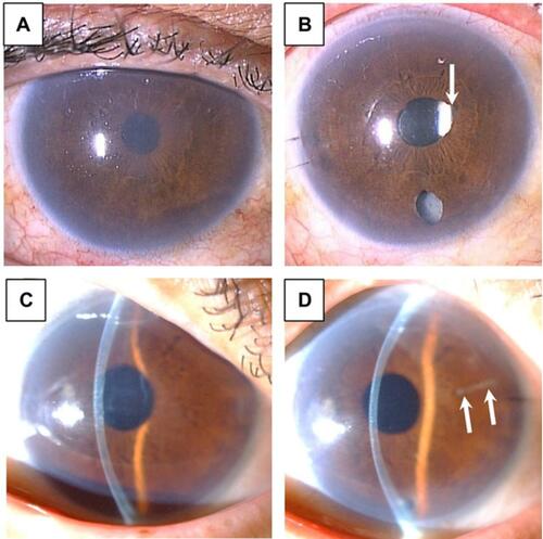
Table 2 Comparison between Group A (with surgery-induced iris abnormalities) and Group B (without surgery-induced iris abnormalities)