Figures & data
Figure 1 Left eye of a 33-year-old man with Vogt-Koyanagi-Harada disease (case 1). (A) Horizontal spectral-domain optical coherence tomography (SD-OCT) image of the fovea before steroid treatment. bar =200 μm. (B, C) SD-OCT image at 4 weeks after steroid treatment when the serous retinal detachment was resolved. Elevation of the RPE layer was observed (arrows). bar =200 μm. Color fundus photograph (D), blue-light fundus autofluorescence (BL-AF) (E), near-infrared fundus autofluorescence (NIR-AF) (F) images of the fundus at the time the serous retinal detachment resolved. In NIR-AF image, hyper autofluorescence in the macula. (G) The AO panorama image of fovea (bar =500 μm). Hyper-reflective lesions were observed at the nasal side of the fovea, and that lesion is corresponding to the elevation of the RPE layer in the OCT. Enlarged images of NIR-AF (H) and AO image (I) of the fovea (image size 2.2×1.1 mm). In the AO image, the boundary of the lesion was clearer. bar =200 μm.
Abbreviations: RPE, retinal pigment epithelium; AO, adaptive optics.
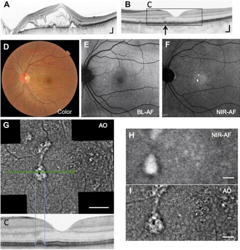
Figure 2 Right eye of a 43-year-old man with Vogt-Koyanagi-Harada disease (case 2). (A) Horizontal spectral-domain optical coherence tomography (SD-OCT) image of the fovea before steroid treatment. bar =200 μm. (B, C) SD-OCT image at 18 months from baseline. Elevation and thickening of the RPE layer were observed near the fovea (arrows). bar =200 μm. Color fundus photograph (D) Blue-light fundus autofluorescence (BL-AF) (E) Near-infrared fundus autofluorescence (NIR-AF) (F) images of the fundus at 18 months from baseline. Slightly hyper-autofluorescence in the BL-AF and hyper-autofluorescence in NIR-AF image was observed. (G) The AO panorama image of fovea (bar =500 μm). Hyper-reflective lesions in AO images are corresponding to the elevation or thinking of the RPE layer in the OCT. Enlarged images of NIR-AF (H) and AO image (I) of the fovea (image size 2.2×1.1 mm). In the AO image, the boundary of the lesion was clearer. bar =200 μm.
Abbreviations: RPE, retinal pigment epithelium; AO, adaptive optics.
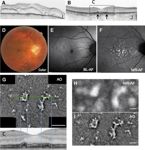
Figure 3 Horizontal SD-OCT images of the fovea from the time when the serous retinal detachment resolved to the final visit. Lesions involving elevation or thickening of the RPE layer were partly diminished (arrows).
Abbreviations: RPE, retinal pigment epithelium; SD-OCT, spectral- domain optical coherence tomography.
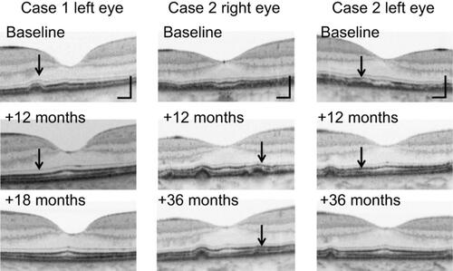
Figure 4 Changes in the adaptive optics (AO) image in the macula of the left eye in case 2. The area of the hyper-reflective lesion in the AO image gradually decreased. All AO image sizes are 2.2×1.1 mm.
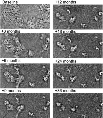
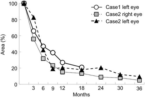
Figure 5 Changes in the area of hyper-reflective lesions in each eyes shown in AO image. The hyper-reflective lesions in the AO image decreased an approximately 40% at 6 months from baseline. However, the hyper-reflective lesions remained to some extent after 18 months in case 1 and 36 months in case 2.
Abbreviation: AO, adaptive optics.