Figures & data
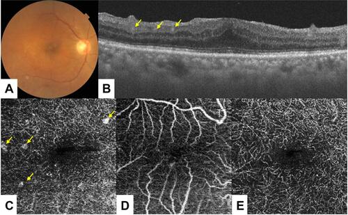
Figure 1 A 74-year-old woman presented with a six-month history of ocular sarcoidosis. Fundus examination of the right eye was unremarkable (A). On spectral-domain OCT and 3×3 mm macular cube OCT angiography conducted in the right eye, however, granulomatous-like lesions were evident in the deeper retinal plexus (B, C: yellow arrow), but not in the superficial plexus (D). After systemic corticosteroid treatment, these lesions were resolved completely (E).