Figures & data
Figure 1 A diagram of the general structure of an amniotic membrane.
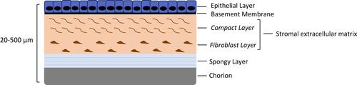
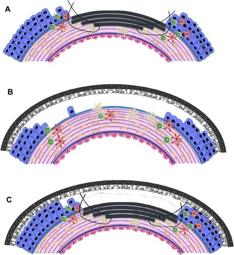
Figure 2 Schematic depicting potential applications and orientation of AMT (depicted in grey) on the ocular surface. (A) Inlay (graft) amnion transplantation. Epithelial-side-up: amnion replaces lost stromal tissue, up to the basement membrane. (B) Onlay (patch) transplantation where amnion is placed epithelial-side-down over the wound periphery as a temporary biological dressing. (C) Combinatorial/Sandwich AMT.