Figures & data
Figure 1 Analysis of Descemet’s membrane perforation types by percentage.
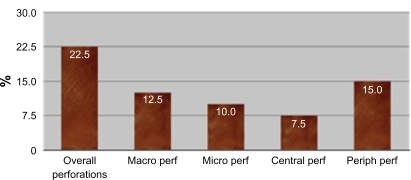
Figure 2 Step at which the perforation occurred.
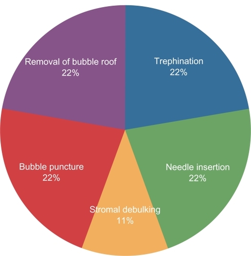
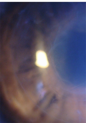
Figure 3 Central preforation.
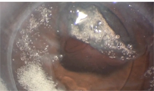
Figure 4 Retained recipient stroma.
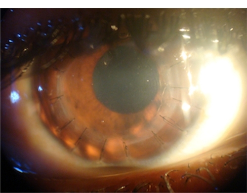
Figure 5 Paracentral perforation with double anterior chamber.
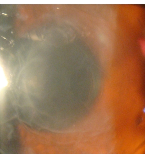
Figure 6 Postoperative Descemet’s membrane wrinkles.