Figures & data

Figure 1 (A) At presentation. Fundus photograph of optic disc, maculoschisis (outlined with white arrows), and inferior coloboma. There is slight depigmentation within the small subfoveal detachment, but no abnormality of the macular retinal pigment epithelium (RPE) elsewhere, since schisis fluid is not in contact with the RPE. (B) Composite OCT topography image of a “giant” maculoschisis cavity, in direct contact with the ODP. (C) OCT cross-section image showing a wide-open pit-macula communication (PMC) (12/15/14, CMT 906 microns, Volume 20.8 mm3).
Abbreviations: OCT, optical coherence tomography; ODP, optic disc pit; CMT, central macular thickness.
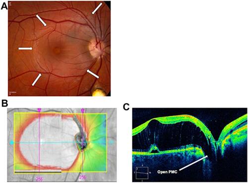
Figure 2 Six weeks after vitrectomy and C3F8 tamponade. (A) Composite OCT topography. (B) OCT cross-section image showing pit-macula communication closed (4/8/15, CMT 501 microns, Volume 15.3 mm3).
Abbreviations: OCT, optical coherence tomography; CMT, central macular thickness.
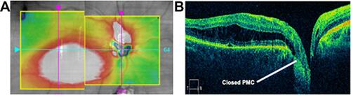
Figure 3 Twelve weeks after vitrectomy and C3F8 tamponade. (A) OCT topographic image showing inferonasal retraction of maculoschisis fluid out of the fovea. (B) OCT cross-section showing sustained closure of the pit-macula communication and resolved foveal schisis (5/27/15, CMT 410 microns, Volume 13.3 mm3).
Abbreviations: OCT, optical coherence tomography; CMT, central macular thickness.
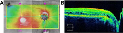
Figure 4 Final follow-up 3.4 years postoperatively. (A) Fundus image showing resolved maculoschisis and closed PMC (subtle optic disc pit is visible inferiorly). (B) Final OCT topographic image of the macula. (C) OCT horizontal section showing a dry, concave fovea with CMT of 322 microns (visual acuity 20/25) (04/02/2018, CMT 322 microns, Volume 10.2 mm3).
Abbreviations: OCT, optical coherence tomography; CMT, central macular thickness; CMT, central macular thickness.
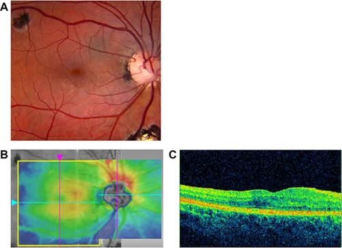
Figure 5 (A) Preoperative OCT showing intact foveal tissue, albeit with schisis and subretinal fluid. (B) OCT image of macular hole that developed after vitrectomy for ODP maculopathy.
Note: Copyright © 2013. Dove Medical Press. Reproduced from Tzu JH, Flynn HW, Berrocal AM, Smiddy WE, Murray TG, Fisher YL. Clinical manifestations of optic pit maculopathy as demonstrated by spectral domain optical coherence tomography. Clin Ophthalmol. 2013;7:167–172.Citation17
Abbreviations: OCT, optical coherence tomography; ODP, optic disc pit.
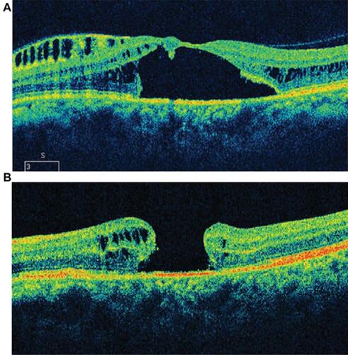
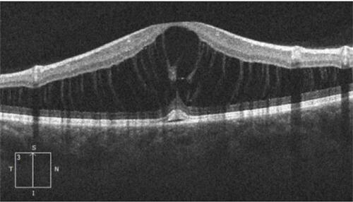
Figure 6 Within the maculoschisis cavity, tenuous axonal strands remain, linking the inner and outer retinal layers.