
Figures & data
Figure 1 Flowchart for inclusion of dry-eye patients to be included in the study to diagnose Sjӧgren’s syndrome.
Abbreviations: TBUT, tear-film breakup time; OSS, ocular staining score; RF, rheumatoid factor; ANA, antinuclear antibody.
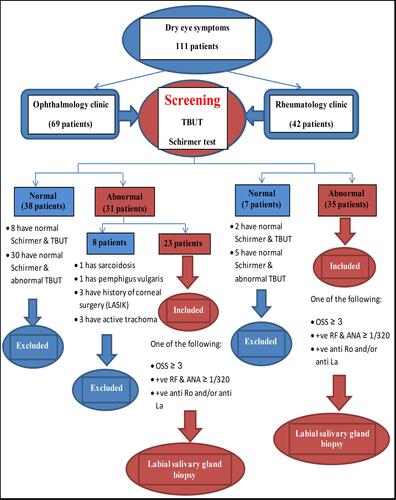
Figure 2 Ocular staining score (OSS). (A) OSS with rose bengal dye giving a score >3. (B) OSS with rose bengal dye giving a score <3.
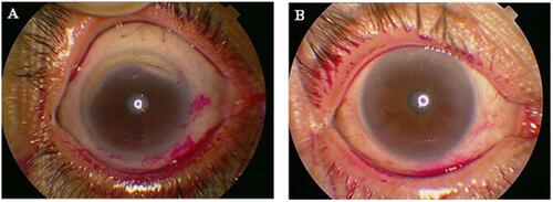
Figure 3 H&E-stained labial salivary glands (LSGs).
Notes: (A) LSGs with three variously sized lymphocytic foci. The entire specimen had a focus score >1/4 mm2 (original magnification 40×). (B) Normal-appearing acini immediately adjacent to the lymphocytic foci (original magnification 100×). (C) LSGs show small lymphocytic aggregate (FLS) that is minimally sized (>50 cells) for inclusion in a focusscore calculation (original magnification 200×).
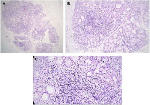
Figure 4 H&E-stained labial salivary glands (LSGs).
Notes: (A) LSGs exhibiting nonspecific chronic sialadenitis with scattered lymphocytes and plasma cells (original magnification 100×). (B) LSGs exhibiting normal glands (original magnification 40×).
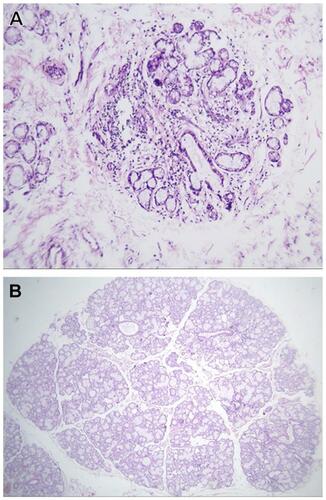
Table 1 Characteristics of dry-eye patients studied
Figure 5 Diagnostic flow of study in Sjӧgren’s syndrome patients.
Abbreviations: OSS, ocular staining score; FLS, focal lymphocytic sialadenitis; NSCS, nonspecific chronic sialadenitis.
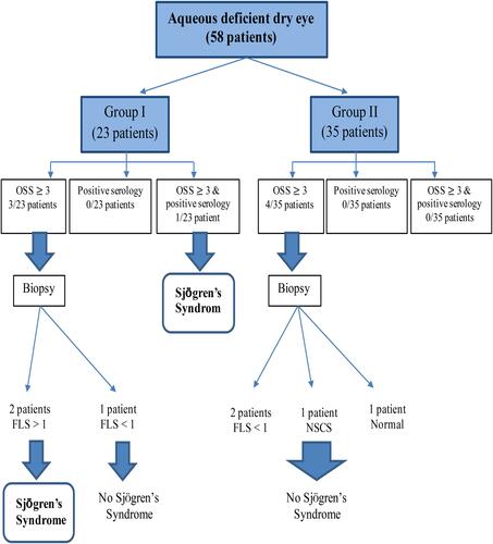
Table 2 Comparisons of ocular tests, autoantibodies, and LSGB with Sjӧgren’s syndrome
Table 3 Sensitivity, specificity, positive predictive value, negative predictive value and accuracy of ocular tests, autoantibodies and LSGB for diagnosis of Sjӧgren’s syndrome
Table 4 Correlations between ocular tests, autoantibodies, LSGB, and Sjӧgren’s syndrome