Figures & data
Table 1 Characteristics of aniridia patients (N=43)
Table 2 Anterior chamber angle and prior surgery in eyes with aniridia (N=86)
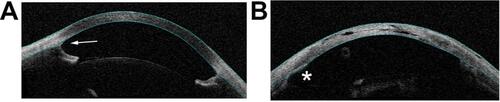
Figure 1 Anterior segment OCT images showing open- and closed-angle findings in aniridia patients. (A) A 12-year-old aniridia patient with glaucoma and open-angle (arrow). (B) A 21-year-old aniridia patient with glaucoma, aphakia, and closed-angle. The asterisk shows an area of angle closure, with iris tissue appositional to the cornea. This patient was treated with lensectomy and goniotomy as an infant, and subsequently with glaucoma drainage implant.