Figures & data
Table 1 Comparison of the Baseline and Final Best-Corrected Visual Acuity (BCVA) and Central Macular Thickness (CMT) After At Least 1 Year of Follow-Up for Eyes with Idiopathic Epiretinal Membranes (iERMs): Group I (VA ≥ 20/30) and Group II (VA 20/40 – 20/50)
Table 2 Changes in the Recorded Visual Acuity and Central Macular Thickness (CMT) in Eyes with Idiopathic Epiretinal Membranes (iERMs) After At Least 1 Year of Follow-Up: Group I (BCVA ≥ 20/30) and Group II (BCVA 20/40 – 20/50)
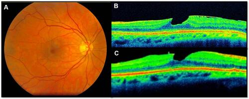
Figure 1 This 70-year-old male patient demonstrates a typical idiopathic epiretinal membrane with central macular involvement and minimal retinal thickening. The initial best-corrected visual acuity (BCVA) was 20/30 and the final BCVA was 20/30. (A) Fundus photograph; (B) baseline OCT image, horizontal cut; (C) final OCT image with wrinkles after 15 months of follow-up.
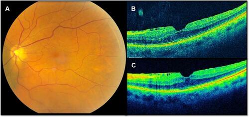
Figure 2 This 58-year-old male demonstrates a typical idiopathic epiretinal membrane with macular involvement and prominent macular thickening. The initial best-corrected visual acuity (BCVA) was 20/40 and the final BCVA was 20/20 after cataract surgery. (A) Fundus photograph; (B) baseline OCT image, horizontal cut; (C) OCT image after 24 months of follow-up.
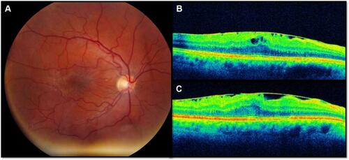
Figure 3 This 62-year-old woman demonstrates a lamellar macular hole. The baseline best-corrected visual acuity (BCVA) was 20/40 and the final BCVA was also 20/40. (A) Fundus photograph; (B) baseline OCT image; (C) OCT image after 24 months follow-up.