Figures & data
Table 1 Preoperative clinical characteristics of the 18 cases
Table 2 Surgical procedures and postoperative clinical characteristics of the 18 cases
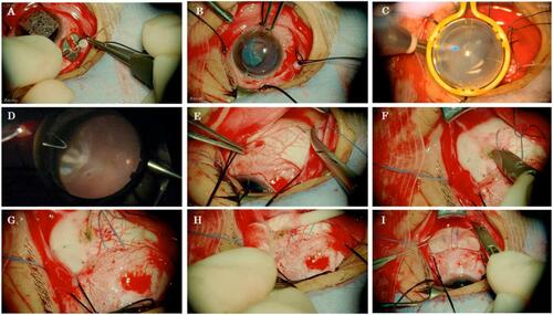
Figure 1 Intraoperative 2D snapshots of chandelier-assisted scleral buckling in case #16. All the images were pictured in the large monitor of the 3D visual system. (A) Following conjunctival peritomy around the limbus and isolation of the rectus muscles with 4–0 silk, a chandelier illumination fiber was placed 4.0 mm behind the limbus at the quadrant opposite the breaks. (B) An anti-drying contact lens was positioned on top of the cornea covered with viscoelastic material. (C) The wide-angle viewing system was activated. (D) Cryopexy was applied to the retinal breaks. With pilot diathermy, flecks applied prior to the chandelier setting were observed. (E) After the light fiber was removed, 5–0 Dacron sutures were applied for the buckle under a microscope with high magnification. (F) Sclerotomy for the external drainage site was also made under a microscope with high magnification. (G) Puncture of the uvea was carried out by a single application of an endolaser. (H) External drainage of subretinal fluid was carried out by scleral depression. (I) The silicone tire was explanted.