
Figures & data
Figure 1 The Visian Toric Implantable Collamer Lens® (VTICL, STAAR Surgical, USA) is available either without (V4b) or with an aquaport (V4c).
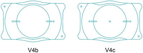
Figure 2 (A) VTICL positioned in the ciliary sulcus as a piggy-back lens. (B) mIOL positioned in the capsular bag which has a circular anterior capsulorhexis.
Abbreviations: mIOL, multifocal intraocular lens; VTICL, Visian Toric Implantable Collamer Lens®.
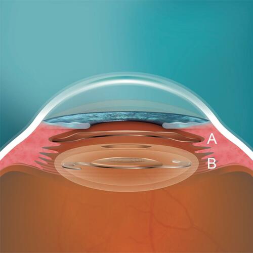
Figure 3 Scheimpflug image (Pentacam® HR, Oculus) from a patient at the 90–270° meridian 3 months after VTICL implantation: (A) distance between endothelium and VTICL: 2400 µm and (B) vault between VTICL and mIOL: 1430 µm.
Abbreviations: mIOL, multifocal intraocular lens; VTICL, Visian Toric Implantable Collamer Lens®.
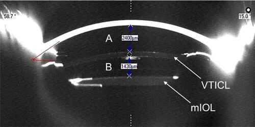
Table 1 Patient demographics and preoperative clinical information
Table 2 Implanted mIOL and Visian Toric Implantable Collamer Lens® models
Table 3 Monocular visual acuity and subjective refraction data before and after VTICL implantation
Figure 4 Cumulative Snellen visual acuity (20/X or better) of pre- and postoperative UDVA.
Abbreviation: UDVA, uncorrected distance visual acuity.
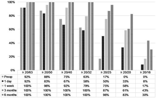
Figure 5 Pre- and postoperative UDVA and CDVA.
Abbreviations: CDVA, corrected distance visual acuity; UDVA, uncorrected distance visual acuity.
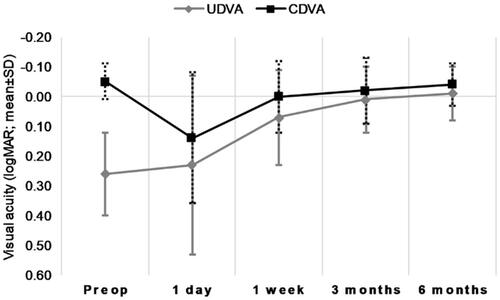
Figure 6 Percentage of eyes achieving the stated accuracy in spherical equivalent.
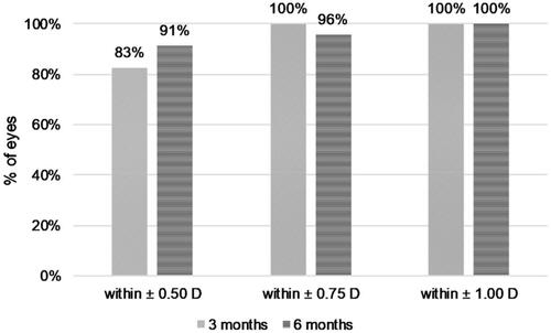
Figure 7 Percentage of eyes achieving the stated accuracy in refractive cylinder.

Figure 8 Distribution of pre-and postoperative refractive astigmatism.
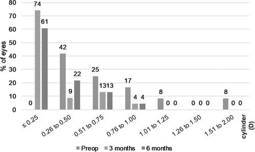
Figure 9 Vector representation (J0 and J45) of refractive astigmatic change after toric ICL implantation. The origin in this graph (0.0) represents an eye free of astigmatism. Abbreviation: ICL, implantable collamer lens.
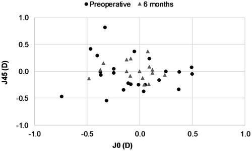
Table 4 Postoperative VTICL misalignment data
Figure 10 Cumulative VTICL misalignment from the target axis over the follow-up period.
Abbreviation: VTICL, Visian Toric Implantable Collamer Lens®.
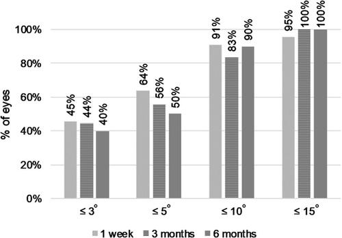
Table 5 Pre- and postoperative ICA data
Table 6 Postoperative clinical outcomes regarding vault (distance between the VTICL and the mIOL) and the distance between the endothelium and the VTICL
Table 7 Preoperative versus 6-month postoperative IOP