Figures & data
Figure 1 A spectral domain optical coherence tomography image of an eye affected by acute central serous chorioretinopathy showing a convex-shaped blister of fluid separating the neurosensory retina from the retinal pigment epithelium and choroid.
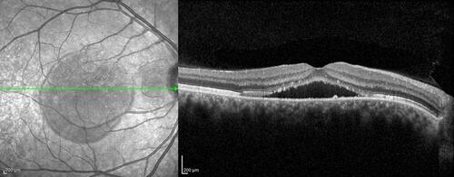
Figure 2 A spectral domain optical coherence tomography image of an eye affected by chronic central serous chorioretinopathy showing a low level of subretinal fluid. At the level of serous detachment, the inner limit is represented by the ellipsoid zone, with elevated photoreceptor tips, and marked reflectivity.
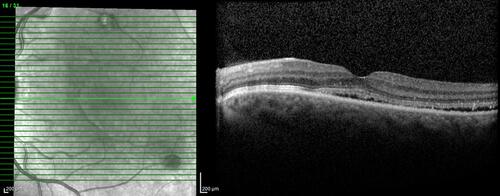
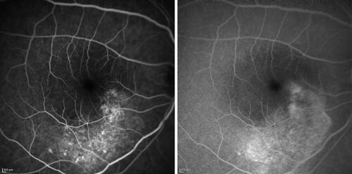
Figure 3 Early and late fluorescein angiograms of a 29-year-old patient affected by chronic central serous chorio-retinopathy revealing an irregularity of the retinal pigment epithelium, resembling a “honeycomb” pattern, near the point of fluorescein leakage. Window defects and late-phase leakage from the expanding point in the superior macula are evident, toward the fovea in a “steam” configuration.