
Figures & data
Figure 1 The wide-angle viewing image is integrated on the large NGENUITY 3D monitor (right) that is placed at the foot of the surgical bed. The 3D monitor for the endoscope (left) is placed 1 m to the front left of the surgeon.
Abbreviation: 3D, three-dimensional.
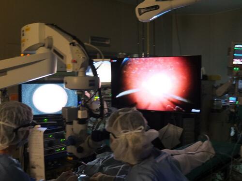
Figure 2 A 3D converter NOVEL HD-3D-A creates 3D endoscopic images in real-time.
Abbreviation: 3D, three-dimensional.
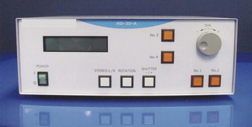
Figure 3 Intraoperative processed 3D endoscope images are recorded as side-by-side stereo images.
Notes: (A) Peripheral vitreous shaving around a retinal break was performed while using the endoscopic view without any scleral depression. (B) Silicone oil was removed through the cannula. Emulsified silicone oil was remarkable.
Abbreviation: 3D, three-dimensional.
