
Figures & data
Figure 1 Diagram showing modified barrier laser around the retinal break and fortified along the vitreous base.
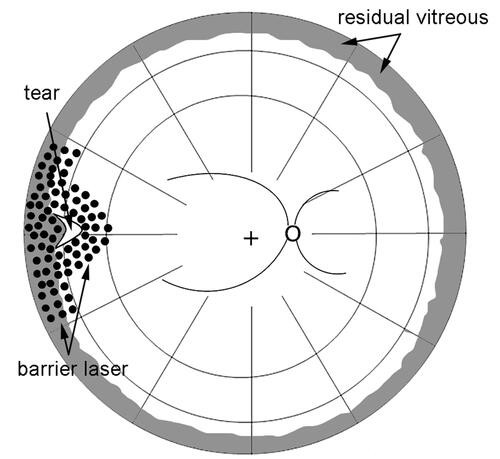
Figure 2 A case of fortified barrier laser for rhegmatogenous retinal detachment (RRD). (A) Ultra-wide field fundus photograph demonstrates RRD in 63-year-old woman with a horseshoe tear and a small atrophic hole. (B) Fortified barrier laser was performed during vitrectomy surrounding two breaks. The flap of the horseshoe tear was removed. (C) The retina was maintained attached after resolution of the tamponade gas.
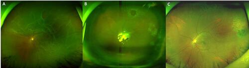
Figure 3 A case of fortified barrier laser for rhegmatogenous retinal detachment (RRD) caused by lattice degeneration. (A) Large tear with lattice degeneration is noted in ultra-wide field fundus photograph. (B) Barrier laser was performed surrounding the tear and lattice degeneration and was fortified along the posterior border of the vitreous base. (C) The reattachment of the retina was maintained after resorption of the gas.
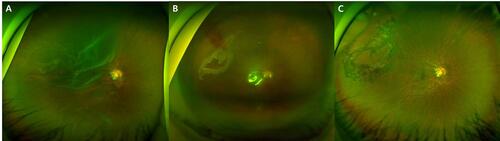
Table 1 Demographics Of The Fortified And Conventional Barrier Laser Groups
Table 2 Clinical Outcomes Of Primary Vitrectomy With Fortified And Conventional Barrier Laser For Rhegmatogenous Retinal Detachment
Table 3 Summary Of Eyes With Recurrent Cases After Primary Vitrectomy For Rhegmatogenous Retinal Detachment