Figures & data
Figure 1 (A) Closed iridocorneal angle observed with a slit lamp. (B) Closed iridocorneal angle observed by optical coherence tomography (OCT). (C) Open iridocorneal angle after phacoemulsification observed with a slit lamp. (D) Open iridocorneal angles after phacoemulsification observed by OCT.
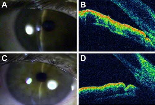
Figure 2 Eyes subjected to phacoemulsification. (A) After glaucomatous crisis. (B) As prophylaxis for glaucoma. (C) After iridotomy.
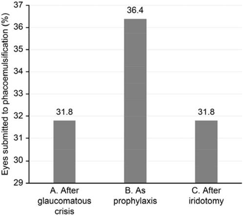
Figure 3 Use of antiglaucomatous medication prior to crisis. (A) Eyes in use of medication. (B) Eyes without use of medication.
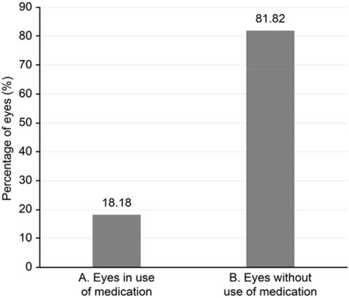
Figure 4 Average intraocular pressure levels after iridotomy and phacoemulsification. (A) Average after iridotomy. (B) Average after phacoemulsification.
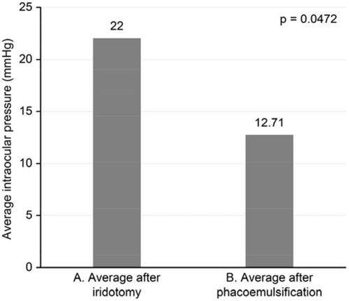
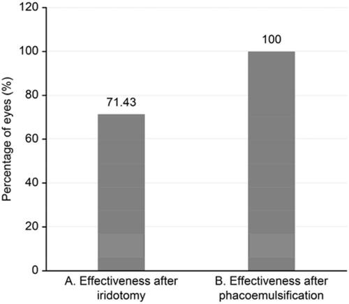
Figure 5 Effectiveness of iridotomy and phacoemulsification in preventing a glaucomatous crisis (minimum of 9-month follow-up). (A) Effectiveness after iridotomy. (B) Effectiveness after phacoemulsification.