Figures & data
Figure 1 Retinal nerve fiber layer (RNFL) thickness (μm) at optic nerve head as compared to best-corrected visual acuity in subjects with unilateral and bilateral optic nerve hypoplasia.
Table 1 Characteristics of Subjects
Table 2 Evaluation of Relationship Between RNFL Thickness of the Optic Nerve Head, as Determined by OCT Spectralis, for Subjects with Either Unilateral or Bilateral ONH, and Recorded Best-Corrected Visual Acuity
Table 3 Summary of Subjects with Nystagmus, Strabismus at Most Recent Office Visit, and Endocrinopathies According to the Laterality of the Optic Nerve Hypoplasia
Table 4 Estimation of the Effects of RNFL Thickness at Optic Nerve Head Domains in the Presence of Nystagmus
Table 5 Estimation of the Effects of RNFL Thickness at Optic Nerve Head Domains in the Presence of Nystagmus
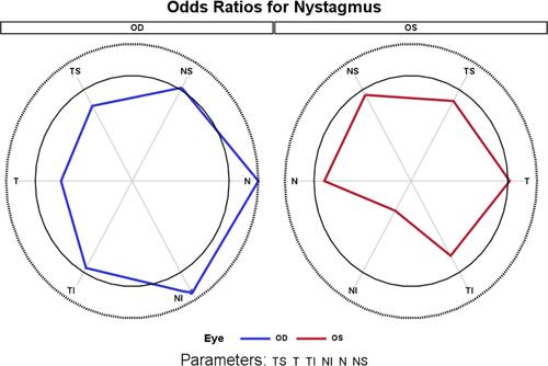
Figure 2 Radar graph depiction of the odds ratio of having nystagmus evaluated at individual optic nerve head regions. In each panel, the outer gray circle represents the maximum value across all the points; the black circle represents an odds ratio of 1 (i.e., no difference). Values within the black circle have an odds ratio of less than 1. Right eye: OD; Left eye: OS. Optic nerve head regions include temporal-superior (TS), temporal (T), temporal-inferior (TI), nasal-inferior (NI), nasal (N), and nasal-superior (NS).